
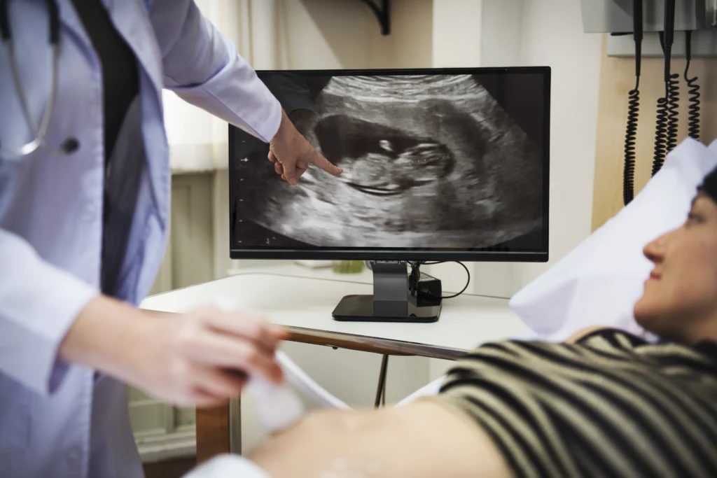
For many expectant parents, the 20-week anatomy scan is one of the most exciting milestones of pregnancy.
You get another opportunity to see your baby.
You may watch tiny movements on the screen.
You might even discover whether you’re having a boy or a girl if you choose to find out.
Quick Answer
The 20-week anatomy scan is a detailed ultrasound performed between 18 and 22 weeks of pregnancy. Doctors examine the baby’s brain, heart, spine, face, kidneys, limbs, placenta, and amniotic fluid to assess growth and development. The scan can identify many structural abnormalities, but it cannot detect every medical condition before birth.
But behind the excitement lies an important medical purpose.
The 20-week anatomy scan is one of the most detailed ultrasound examinations performed during pregnancy.
It helps healthcare providers assess how your baby is developing and identify many structural abnormalities before birth.
While most scans provide reassuring news,
understanding what doctors are actually looking for can help you approach the appointment with confidence and realistic expectations.
Key Takeaways
- The 20-week anatomy scan is one of the most important pregnancy ultrasounds.
- It is usually performed between 18 and 22 weeks of pregnancy.
- Doctors examine the baby’s brain, heart, spine, organs, and limbs.
- The scan also evaluates placental position and amniotic fluid levels.
- Many structural abnormalities can be detected before birth.
- A normal anatomy scan is reassuring but cannot guarantee perfect health.
- Soft markers may sometimes be identified and require further assessment.
- Additional testing does not automatically mean there is a serious problem.
- Most anatomy scans are normal and reassuring.
- The scan helps healthcare providers plan the best possible care for mother and baby.
What Is The 20-Week Anatomy Scan?
The 20-week anatomy scan is a detailed ultrasound examination usually performed between 18 and 22 weeks of pregnancy.
You may also hear it called:
- Anatomy scan
- Mid-pregnancy scan
- Detailed fetal anatomy scan
- Level II ultrasound
Unlike early pregnancy scans that focus on confirming pregnancy or estimating the due date, this examination evaluates the baby’s anatomy in a systematic and detailed manner.
Doctors use the scan to examine major organs, body systems, growth patterns, and the pregnancy environment. (Bethune et al. 2015)
The second-trimester ultrasound is considered one of the most comprehensive fetal assessments performed during routine prenatal care.
Why Is The 20-Week Scan So Important?
By the middle of pregnancy, most major organs have developed sufficiently to be examined in detail.
This timing allows healthcare providers to evaluate structures that may not have been visible during earlier scans.
The anatomy scan plays a central role in modern prenatal care because it can:
- Assess fetal growth
- Evaluate organ development
- Detect many structural abnormalities
- Examine placental location
- Assess amniotic fluid volume
- Guide pregnancy management when necessary
Research has shown that structured prenatal screening programs improve the ability to identify fetal conditions before birth and support appropriate pregnancy care planning. (Wilson et al. 2024)
The First Thing Doctors Check: Growth And Measurements
One of the earliest steps during the anatomy scan is assessing whether the baby appears to be growing appropriately.
Several measurements are obtained, including:
- Head circumference
- Biparietal diameter (head width)
- Abdominal circumference
- Femur length
These measurements help estimate fetal growth and determine whether development appears consistent with gestational age.
Growth assessment remains an essential component of second-trimester ultrasound examinations. (Bethune et al. 2015)
Looking Closely At The Baby’s Brain
The brain is one of the most carefully evaluated structures during the anatomy scan.
Doctors assess:
- Skull shape
- Brain symmetry
- Ventricular system
- Midline structures
- Posterior fossa
These observations help determine whether brain development appears appropriate for gestational age.
Because the fetal nervous system continues developing throughout pregnancy, some findings may require follow-up imaging later.
Examining The Face
The baby’s face is also evaluated during the scan.
This assessment may include:
- Facial profile
- Nose
- Lips
- Eye positioning
The purpose is not cosmetic.
Instead, certain facial findings may occasionally provide valuable information about fetal development and potential structural abnormalities.
The Heart: One Of The Most Important Parts Of The Scan
Congenital heart defects are among the most common birth abnormalities.
For this reason, the fetal heart receives special attention during the anatomy scan.
The sonographer evaluates:
- Heart position
- Four cardiac chambers
- Major blood vessels
- Cardiac outflow tracts
Obtaining high-quality cardiac views is one of the most important goals of the examination.
If concerns arise, a fetal echocardiogram may be recommended for a more detailed assessment.
Evaluating The Spine
The spine is examined from multiple angles to assess continuity and development.
This evaluation helps healthcare providers identify abnormalities involving the spinal cord and surrounding structures.
Conditions such as certain neural tube defects may sometimes be identified through detailed ultrasound assessment.
Checking The Abdomen And Internal Organs
Several internal organs are evaluated during the scan.
These include:
Stomach
Doctors confirm the stomach can be visualized and appears appropriately positioned.
Kidneys
Both kidneys are assessed for development and appearance.
Bladder
The bladder is examined as part of the urinary system evaluation.
Abdominal Wall
The abdominal wall is assessed to ensure internal organs appear appropriately enclosed.
This part of the examination helps identify abnormalities affecting multiple body systems.
Looking At The Arms, Legs, Hands, And Feet
The anatomy scan also evaluates:
- Arms
- Legs
- Hands
- Feet
- Long bones
The goal is to assess development, movement, and overall growth patterns.
The presence of normal limb movement provides additional reassurance regarding fetal well-being.
Placenta And Amniotic Fluid Assessment
The anatomy scan does not focus exclusively on the baby.
Doctors also examine the pregnancy environment.
Placental Position
The placenta’s location is documented because it may influence pregnancy management later.
Amniotic Fluid
The amount of amniotic fluid is assessed to ensure the baby has an appropriate environment for growth and development.
What Research Shows About Detection
Modern second-trimester ultrasound examinations can identify many clinically significant fetal abnormalities before birth.
Research demonstrates that routine anatomical assessment during pregnancy improves prenatal detection of structural abnormalities and supports earlier counseling and care planning. (Syngelaki et al. 2019)
A Thought Before Your Scan
Most parents walk into the anatomy scan hoping for a simple answer:
Is my baby okay?
While no test can guarantee perfect health, the 20-week anatomy scan provides one of the most detailed assessments available during pregnancy.
For most families, the appointment offers reassurance, excitement, and a clearer understanding of how their baby is growing.
What Can The 20-Week Anatomy Scan Detect?
One of the biggest reasons the anatomy scan is performed is to identify structural abnormalities before birth.
The scan can help detect certain conditions affecting:
- The brain
- Heart
- Spine
- Kidneys
- Abdomen
- Limbs
- Face
- Urinary system
Some abnormalities may be minor and require only monitoring.
Others may need additional investigations, specialist consultations, or delivery planning.
The purpose of identifying these conditions before birth is not to create anxiety but to ensure families and healthcare providers have the information needed to make informed decisions.
Research has shown that routine second-trimester ultrasound examinations can identify many major non-chromosomal abnormalities before birth when systematic anatomical assessment is performed. (Syngelaki et al. 2019)
What The Anatomy Scan Cannot Detect
This is one of the most important things expectant parents should understand.
The anatomy scan is powerful, but it is not perfect.
A normal anatomy scan does not guarantee that a baby has no medical conditions.
Some conditions:
- Develop later in pregnancy
- Are too subtle to detect on ultrasound
- Involve genetic changes without visible anatomical findings
- May not be detectable before birth
Even the most experienced specialists and the most advanced ultrasound equipment have limitations.
This is why healthcare providers describe the anatomy scan as an assessment tool rather than a guarantee.
What Are Soft Markers?
Sometimes the anatomy scan identifies what doctors call soft markers.
A soft marker is not a birth defect.
Instead, it is an ultrasound finding that may be associated with an increased likelihood of certain chromosomal conditions.
Examples include:
- Echogenic intracardiac focus
- Mild renal pelvic dilation
- Echogenic bowel
- Increased nuchal fold thickness
Importantly, many healthy babies have isolated soft markers.
When one is identified, healthcare providers consider:
- Maternal age
- Previous screening results
- Family history
- Other ultrasound findings
The presence of a soft marker does not automatically mean that a chromosomal condition exists.
Can The Anatomy Scan Detect Down Syndrome?
Many parents assume the anatomy scan can diagnose Down syndrome.
It cannot.
Down syndrome is a chromosomal condition caused by an extra copy of chromosome 21.
While ultrasound may identify certain markers that increase suspicion, the anatomy scan alone cannot confirm the diagnosis.
In some cases, doctors may observe:
- Increased nuchal fold thickness
- Certain cardiac findings
- Specific anatomical variations
These findings may lead to recommendations for additional screening or diagnostic testing.
The anatomy scan contributes information, but it is only one piece of the overall assessment.
What Happens If Doctors Find Something Unexpected?
Hearing that further testing is needed can be stressful.
However, additional testing does not automatically mean something is seriously wrong.
Many follow-up assessments are recommended simply because:
- The baby was not in an ideal position
- Certain structures could not be visualized clearly
- Additional measurements are required
- Clarification is needed
Possible next steps may include:
Repeat Ultrasound
A follow-up scan may provide better images or monitor a finding over time.
Fetal Echocardiography
A detailed assessment of the baby’s heart.
Genetic Counseling
Provides information about potential findings and available testing options.
Diagnostic Testing
In selected situations, procedures such as amniocentesis may be discussed.
The goal is always to gather more information before drawing conclusions.
Why Early Detection Can Be Helpful
Prenatal detection of abnormalities often allows healthcare teams to prepare for delivery and postnatal care.
Depending on the condition identified, healthcare providers may:
- Arrange specialist consultations
- Plan delivery at an appropriate facility
- Coordinate neonatal care
- Monitor the pregnancy more closely
Early detection can improve preparedness and help families understand available options.
Common Myths About The 20-Week Anatomy Scan
Myth 1: The Scan Is Just To Find Out The Baby’s Gender
False.
The primary purpose of the anatomy scan is medical assessment of fetal anatomy and development.
Gender identification is only a small part of the examination.
Myth 2: A Normal Scan Guarantees A Healthy Baby
False.
A normal scan is reassuring but cannot exclude every condition.
Myth 3: Every Abnormal Finding Means A Serious Problem
False.
Many findings turn out to be minor variations or require only monitoring.
Additional testing is often performed simply to gather more information.
Myth 4: The Scan Can Diagnose Every Genetic Disorder
False.
Many genetic conditions cannot be diagnosed through ultrasound alone.
Additional screening or diagnostic testing may be needed.
| Structure Examined | Why It Matters |
|---|---|
| Brain | Assesses neurological development |
| Heart | Screens for congenital heart defects |
| Spine | Evaluates spinal development |
| Face | Assesses facial anatomy |
| Kidneys & Bladder | Evaluates urinary tract development |
| Limbs | Assesses growth and movement |
| Placenta | Checks pregnancy support system |
| Amniotic Fluid | Monitors fetal environment |
Questions Parents Frequently Ask
“How Long Does The Scan Take?”
Most anatomy scans take approximately 20-45 minutes.
The duration depends on fetal position, image quality, and whether additional views are required.
“Can I Eat Before The Scan?”
In most cases, yes.
Your healthcare provider will advise you if any special preparation is needed.
“Will I Get Pictures Of My Baby?”
Many ultrasound centers provide images, although policies vary.
“What If My Baby Is Facing The Wrong Direction?”
This happens frequently.
The sonographer may ask you to move, walk, or change positions to improve visualization.
“Can The Sonographer Tell Me The Results Immediately?”
Practices vary by facility.
Some findings may be discussed during the examination, while others are reviewed with your healthcare provider afterward.
A Note From A Physiotherapist
Many parents place enormous emotional weight on the 20-week scan.
They spend days preparing for the appointment and worrying about every possible outcome.
While those feelings are understandable, it helps to remember why the scan exists.
The anatomy scan is a tool.
Its purpose is to provide information.
Most scans are reassuring.
When concerns are identified, the information allows healthcare teams to provide better support, additional monitoring, and appropriate care.
The scan is not designed to create fear.
It is designed to help.
What The Scan Cannot Tell You About Your Baby
Even the most detailed ultrasound cannot predict:
- Personality
- Intelligence
- Talents
- Interests
- Future achievements
- Emotional development
Parents sometimes forget this because modern technology is so impressive.
Yet every child remains wonderfully unique and unpredictable.
One Less Thing To Worry About
Many women arrive at the anatomy scan convinced they are going to receive bad news.
In reality, most anatomy scans are reassuring.
Most babies show normal growth and development.
Most parents leave the appointment feeling relieved, excited, and more connected to their baby than ever before.
If Nobody Has Told You This Today…
You do not need to understand every ultrasound measurement.
You do not need to memorize medical terminology.
And you certainly do not need to become an expert in fetal medicine.
Your role is simply to attend your appointments, ask questions, and work with your healthcare team.
That is enough.
Final Thoughts
The 20-week anatomy scan is one of the most important ultrasound examinations performed during pregnancy
because it provides a detailed assessment of fetal anatomy, growth, and development.
By examining major organs, body systems, placental position, and the pregnancy environment, healthcare providers can identify many structural abnormalities and plan care appropriately when needed.
Although the scan cannot detect every condition before birth, it remains one of the most valuable tools available in modern prenatal care.
The most important thing to remember is this:
The 20-week anatomy scan is not about finding problems.
It is about understanding how your baby is developing and ensuring that both you and your healthcare team have the information needed to support a healthy pregnancy.
In One Sentence
The 20-week anatomy scan provides one of the most detailed assessments of your baby’s development before birth.
Frequently Asked Questions About The 20-Week Anatomy Scan
What is the 20-week anatomy scan?
The 20-week anatomy scan is a detailed ultrasound examination used to assess fetal growth, anatomy, and development during the second trimester.
When is the anatomy scan performed?
Most anatomy scans are performed between 18 and 22 weeks of pregnancy.
What does the anatomy scan check?
The scan evaluates the baby’s brain, heart, spine, face, kidneys, bladder, limbs, placenta, and amniotic fluid.
Can the anatomy scan detect birth defects?
The scan can identify many structural abnormalities but cannot detect every medical condition before birth.
Can the anatomy scan detect Down syndrome?
The scan may identify markers associated with Down syndrome, but it cannot diagnose the condition.
How long does the scan take?
Most anatomy scans take approximately 20–45 minutes, depending on fetal position and image quality.
What happens if the doctor finds something unusual?
Additional imaging, genetic counseling, fetal echocardiography, or diagnostic testing may be recommended depending on the findings.
Can a normal anatomy scan guarantee a healthy baby?
No. A normal scan is reassuring but cannot rule out every developmental, genetic, or medical condition.
Stay tuned with us for more health related topics.
Follow us on LinkedIn and Instagram for more.
More Read


Medical Disclaimer!
This article has been reviewed and written under the guidance of our Head Physiotherapist, Dr. Kruti Raj (PT, MUHS,CPT,CMPT). The information shared is intended for educational purposes only and should not be considered a substitute for personalized medical advice, diagnosis, or treatment.
Please consult us or any other qualified healthcare professional before beginning any exercise program, especially if you are experiencing pain, recovering from injury, or managing a medical condition.


