


An ankle sprain usually improves gradually, but persistent deep ankle pain, swelling or discomfort during walking may indicate an injury beyond the ligaments.
Quick Answer: Why Does My Ankle Still Hurt?
Persistent ankle pain after a sprain may sometimes indicate an osteochondral lesion of the talus (OLT). This injury affects the cartilage covering the talus and the underlying bone, and it can occur alongside a ligament sprain.
Deep ankle pain, recurring swelling, stiffness or catching during movement may warrant further assessment, especially when recovery is not progressing as expected.
What should you do? Seek clinical assessment for persistent symptoms. Depending on the findings, MRI or CT may be needed. Treatment can include protected activity, individualised physiotherapy or surgery for selected lesions.
One possible cause is an osteochondral lesion of the talus, which affects the smooth cartilage covering the ankle bone and the underlying bone tissue.
This injury can be difficult to recognize because its symptoms may resemble an ordinary sprain.
Understanding the warning signs, diagnostic tests and role of physiotherapy can help people seek appropriate treatment before returning to demanding activities.
Key Takeaways About Osteochondral Lesions
Cartilage and bone can both be affected. An OLT involves the joint surface of the talus and the bone beneath it.
A sprain does not rule out another injury. Persistent deep pain, recurring swelling or catching may warrant further investigation.
A normal X-ray may not be enough. MRI or CT may be considered when symptoms and examination findings suggest an osteochondral lesion.
Rehabilitation must match the injury. Joint protection, progressive exercises and return-to-sport planning should follow the treatment team’s guidance.
What Is an Osteochondral Lesion of the Talus?
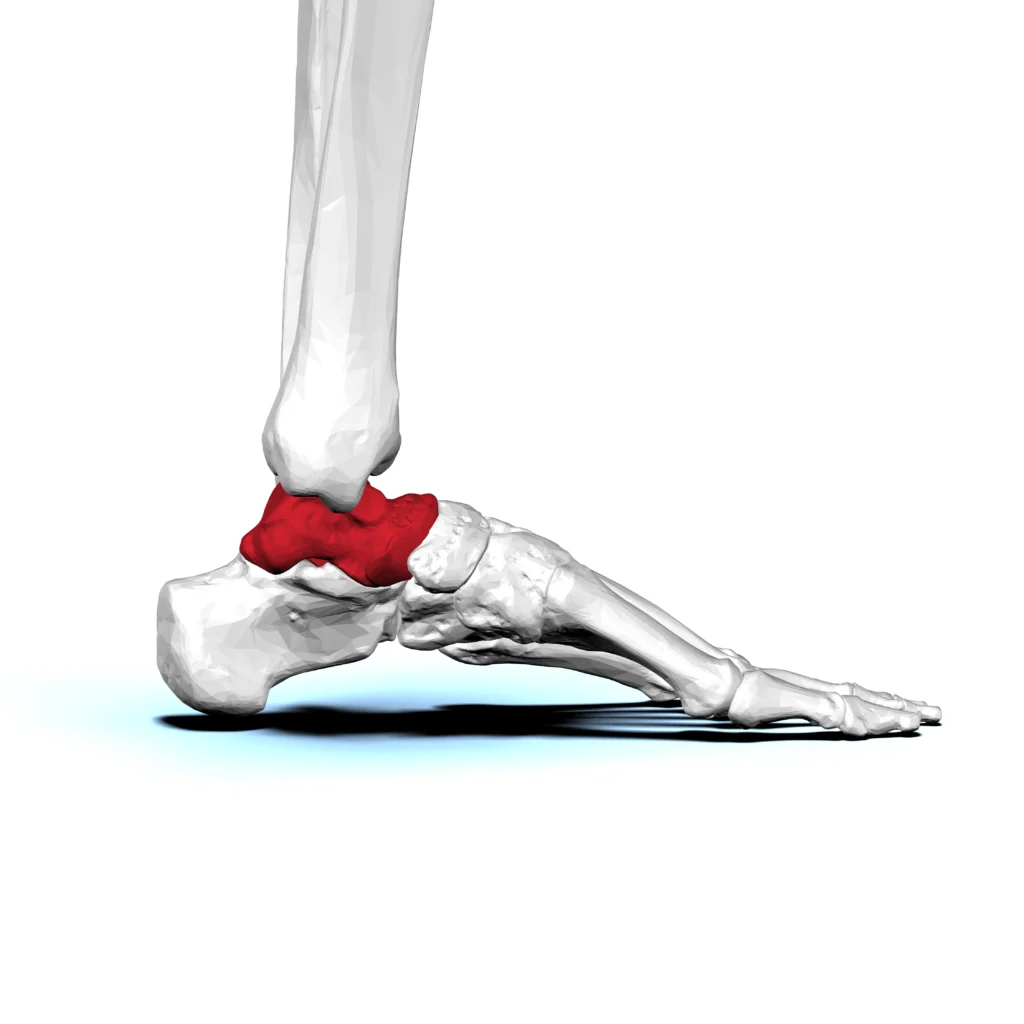
The talus is the bone that connects the lower leg to the foot and helps form the ankle joint.
Its upper surface, called the talar dome, is covered with articular cartilage that allows the joint to move smoothly.
An osteochondral lesion of the talus (OLT) is an injury or abnormality involving this cartilage and the bone immediately beneath it.
The lesion may range from damage within the bone to cartilage disruption or a partially or completely detached fragment.
OLT can develop after a twisting injury, ankle sprain or fracture.
Some lesions also occur without a clearly identifiable traumatic event.
Anastasio et al. explain that treatment depends on factors such as lesion size, stability, location and the patient’s symptoms. (Anastasio et al., 2024)
Why Does Your Ankle Still Hurt After a Sprain?
A typical ankle sprain affects the ligaments that support the joint.
However, during a forceful twist, the talus may also strike the surrounding bones, damaging its cartilage and underlying bone.
This can happen during an inversion injury, when the foot rolls inward.
The cartilage may become damaged even when the initial X-ray does not reveal a fracture.
An osteochondral lesion may therefore explain why someone continues to experience pain after the visible swelling and bruising from a sprain have improved.
Persistent pain does not automatically mean an OLT is present.
Other possibilities include an incompletely healed ligament injury, tendon damage, joint irritation or an occult fracture.
A clinical assessment is needed to distinguish them.
Symptoms of an Osteochondral Lesion of the Talus
Symptoms vary according to the size, location and stability of the lesion.
Some lesions cause few symptoms, while others interfere significantly with walking and exercise.
Deep ankle pain during activity
Pain may feel as though it comes from inside the joint rather than from the skin or surrounding ligaments.
It may worsen during walking, running, jumping or other weight-bearing activities.
Persistent or recurring swelling
The ankle may swell after exercise or prolonged standing.
Swelling that repeatedly returns despite appropriate recovery from a sprain deserves assessment.
Stiffness and reduced movement
The ankle may feel stiff when bending forward, squatting or moving through its full range.
Pain and joint irritation can make everyday movements uncomfortable.
Catching, clicking or locking
Some people experience catching or a sensation that the ankle briefly gets stuck.
This can occur when a cartilage or bone fragment is unstable, although clicking alone does not confirm an osteochondral lesion.
Pain that returns when activity increases
A person may feel better during rest but notice pain again when returning to running, sports or longer walks.
Repeatedly increasing activity without understanding the cause can prolong symptoms.
How Is an OLT Different From a Normal Ankle Sprain?
| Feature | Typical ankle sprain | Possible osteochondral lesion |
|---|---|---|
| Main structure affected | Supporting ligaments | Joint cartilage and underlying bone |
| Pain location | Often around the outer ankle after an inversion sprain | May feel deeper inside the joint |
| Swelling | Usually more noticeable early after injury | May persist or recur with activity |
| Recovery pattern | Often improves progressively with rehabilitation | May remain painful despite routine sprain rehabilitation |
| Additional symptoms | Ligament tenderness and instability | Possible stiffness, catching or locking |
These patterns can overlap.
An OLT and a ligament sprain can occur together, so persistent symptoms should not be diagnosed from a comparison table alone.
Osteochondral Lesion of the Talus: Myth vs Fact
MYTH: Persistent pain after a sprain is always caused by damaged ligaments.
FACT: Cartilage and underlying bone injuries, tendon problems and occult fractures can also cause ongoing ankle pain.
MYTH: A normal X-ray rules out an osteochondral lesion.
FACT: Some lesions are not clearly visible on plain X-rays. Further imaging may be appropriate when symptoms remain suspicious.
MYTH: Every osteochondral lesion requires surgery.
FACT: Some stable lesions may be managed conservatively, while others need surgery based on their characteristics and symptoms.
MYTH: If ankle pain improves, returning to sport immediately is safe.
FACT: Return to sport should account for lesion stability, prescribed loading restrictions, strength, movement and clinical clearance where required.
How Is an Osteochondral Lesion Diagnosed?
Diagnosis involves a clinical examination and, when indicated, imaging.
The clinician will ask about the original injury, symptom progression, activity-related pain and any episodes of catching or locking.
Physical examination
The examination may include ankle range of motion, swelling, tenderness, ligament stability, walking ability and functional limitations.
These findings help identify possible causes but cannot reliably confirm or exclude every cartilage injury.
X-rays
X-rays are often the initial imaging test after an ankle injury.
They can identify fractures and some osteochondral abnormalities.
However, small or early lesions may not be visible on plain radiographs.
A normal X-ray does not necessarily rule out an OLT when symptoms remain suspicious.
MRI and CT scans
Magnetic resonance imaging (MRI) can assess cartilage, underlying bone changes, bone marrow oedema and associated soft-tissue injuries.
It is particularly useful when an osteochondral lesion is suspected despite normal X-rays.
Computed tomography (CT) provides detailed information about bone structure, lesion dimensions and cysts, and may help with surgical planning.
Khan et al. review the role of these imaging methods in identifying and staging talar osteochondral lesions. (Khan et al., 2024)
Treatment Options for an Osteochondral Lesion
Treatment depends on the lesion’s size, stability, symptoms, activity demands and response to initial management.
Not every lesion requires surgery.
Conservative treatment
Selected stable or nondisplaced lesions may initially be managed without surgery.
Depending on the injury, the plan may include temporary activity restriction, protected weight-bearing or immobilization under medical supervision.
The aim is to avoid excessive joint loading while the lesion is assessed and symptoms settle.
The appropriate duration of protection varies, so patients should not independently continue sports or force weight-bearing through pain.
Walther et al.’s recommendations highlight activity modification as an important part of conservative management, while noting that outcomes vary and follow-up may be necessary. (Walther et al., 2023)
Surgical treatment
Surgery may be considered when symptoms persist despite appropriate non-operative treatment or when the lesion has features that make conservative care unsuitable.
A displaced fragment may require earlier specialist intervention.
Depending on the lesion, procedures may involve removing unstable tissue, stimulating repair of the underlying bone, fixing a suitable fragment or using cartilage and bone restoration techniques.
There is no single operation appropriate for every OLT.
The orthopedic specialist considers the lesion’s characteristics and the person’s functional needs when selecting treatment.
Physiotherapy for an Osteochondral Lesion of the Talus
Physiotherapy is important for restoring ankle function, but its timing and intensity must match the lesion and the treatment plan.
An osteochondral injury should not automatically be managed like an uncomplicated ankle sprain.
Protect the joint during early recovery
When the lesion requires restricted loading or immobilization, the physiotherapist works within the treating clinician’s instructions.
Weight-bearing should not be increased simply because pain has temporarily improved.
Education about activity modification, safe walking and avoiding aggravating movements can help protect the joint during this stage.
Restore ankle movement when cleared
Once appropriate, rehabilitation may include gentle range-of-motion exercises to address stiffness and restore comfortable movement.
The timing depends on lesion stability, healing requirements and whether surgery was performed.
Exercises should not force painful movement or conflict with prescribed restrictions.
Rebuild strength and balance
As recovery progresses, the program may include progressive strengthening of the calf and ankle muscles, balance exercises and walking retraining.
These exercises help address weakness and movement deficits that can remain after an ankle injury.
Progression should be based on symptoms, function and clinical guidance rather than a fixed timeline.
Prepare for running and sport
Returning to running, jumping or cutting movements requires more than reduced pain.
Rehabilitation may need to address walking tolerance, ankle mobility, strength, balance and sport-specific demands.
A graded return is appropriate only when permitted by the treating team.
Persistent deep pain, swelling or catching during progression should prompt reassessment.
How Long Does Recovery Take?
Recovery varies considerably.
It depends on lesion size and stability, the severity of the original injury, the treatment chosen and individual healing factors.
A mild, stable lesion managed conservatively may improve over time, while a more substantial lesion or one requiring surgery may need a longer period of protected activity and rehabilitation.
Buck et al.’s systematic review found that non-operative outcomes varied considerably across studies, with an overall pooled clinical success rate of 45%.
The studies differed in treatment methods and patient characteristics, so this figure cannot predict an individual’s recovery. (Buck et al., 2023)
Do not use pain alone to decide when to resume sport.
Follow-up assessment and, in selected cases, repeat imaging may be needed before returning to high-impact activity.
When Should You Seek Medical Attention?
Arrange an assessment if ankle pain persists or repeatedly returns after a sprain, particularly if it is accompanied by recurring swelling, stiffness or catching inside the joint.
Seek urgent medical assessment if you cannot bear weight after a new injury, develop severe or rapidly worsening pain and swelling, or notice a visibly deformed ankle.
A hot, red joint accompanied by fever also needs urgent evaluation.
A clinician can determine whether imaging or referral to an orthopedic specialist is appropriate.
Lesser-Known Facts About Osteochondral Lesions
Cartilage damage may not appear on an X-ray
Early cartilage and underlying bone changes can be missed on plain radiographs.
Persistent symptoms may justify further investigation even when the initial X-ray is normal.
An OLT can exist alongside a ligament injury
Finding a sprain does not exclude an additional cartilage or bone injury.
When recovery does not follow the expected pattern, the original diagnosis may need to be reviewed.
Symptoms and scan findings do not always match
Some lesions are discovered incidentally, while others cause substantial pain.
Clinicians consider the imaging findings alongside symptoms, examination and functional limitations rather than treating a scan result in isolation.
Final Thoughts: Persistent Ankle Pain Deserves Attention
An osteochondral lesion of the talus is one possible reason ankle pain continues after a sprain. Deep joint pain, recurring swelling, stiffness or catching may indicate an injury that needs further assessment.
Appropriate imaging and individualised treatment can help guide recovery. Physiotherapy supports movement and function, but loading and return to sport should follow the lesion-specific treatment plan.
Conclusion
An osteochondral lesion of the talus is an important possible cause of persistent ankle pain after a sprain.
Damage to the joint cartilage and underlying bone may produce deep pain, recurring swelling, stiffness or catching that does not improve as expected with routine sprain rehabilitation.
Appropriate assessment and imaging can help identify the lesion and guide treatment.
Physiotherapy supports recovery through joint protection, carefully timed movement, progressive strengthening and a planned return to activity, while the need for surgery depends on the lesion and its response to conservative care.
Frequently Asked Questions
1. Can an ankle sprain cause an osteochondral lesion?
Yes. A twisting injury can damage the cartilage and underlying bone of the talus. An osteochondral lesion may occur alongside a ligament sprain.
2. Can an osteochondral lesion heal without surgery?
Some stable lesions improve with appropriate conservative management. Others remain symptomatic or have features that require surgery. Treatment depends on the lesion and clinical assessment.
3. Why does my ankle still hurt months after a sprain?
Persistent pain may result from ligament injury, tendon damage, an occult fracture or an osteochondral lesion. Continued pain deserves assessment rather than being automatically attributed to slow healing.
4. Is walking safe with an osteochondral lesion?
It depends on the lesion’s stability and treatment plan. Some people can walk with modified activity, while others need protected weight-bearing or immobilisation under medical guidance.
5. Can physiotherapy cure an osteochondral lesion?
Physiotherapy can support mobility, strength, balance and functional recovery, but it cannot guarantee cartilage or bone healing. Some lesions need additional medical or surgical treatment.
Stay tuned with us for more health related topics.
Follow us on LinkedIn and Instagram for more.
More Read


Medical Disclaimer
This article is intended for general educational purposes and does not replace professional medical advice, diagnosis or treatment. Persistent ankle pain after a sprain can have several causes, and symptoms alone cannot confirm an osteochondral lesion.
Consult a qualified healthcare professional for assessment. Seek urgent care for severe or rapidly worsening symptoms, ankle deformity, or inability to bear weight after a new injury.


