


Advanced fetal imaging during pregnancy gives doctors a more detailed view of :
your baby’s growth, anatomy, and well-being, helping identify certain conditions that routine scans may not fully evaluate.
Pregnancy is an exciting journey filled with milestones, and one of the most reassuring moments is seeing your baby during an ultrasound scan.
Advances in medical imaging have transformed prenatal care, allowing healthcare professionals to assess fetal growth, anatomy, organ development, and blood flow with greater accuracy than ever before.
While most pregnancies are monitored using routine ultrasound examinations, some situations require advanced fetal imaging techniques to obtain additional information.
These specialized imaging methods help doctors evaluate suspected:
congenital abnormalities, monitor high-risk pregnancies, assess fetal heart function, examine blood circulation, and better understand complex conditions before birth.
Quick Answer
Advanced fetal imaging techniques provide detailed information about your baby’s development when routine pregnancy ultrasound requires further evaluation. These techniques include high-resolution ultrasound, Doppler ultrasound, fetal echocardiography, 3D ultrasound, 4D ultrasound, and fetal MRI. Most pregnancies do not require every imaging test, and your healthcare provider will recommend the most appropriate examination based on your individual pregnancy.
Many expecting parents become anxious when their healthcare provider recommends an additional scan.
It is natural to wonder whether something is wrong or whether advanced imaging means the pregnancy has become high risk.
In reality, these tests are often performed to gather more detailed information, confirm normal findings, or guide the most appropriate care for both mother and baby.
Modern fetal imaging now includes several specialized techniques such as:
- high-resolution ultrasound,
- Doppler ultrasound,
- 3D ultrasound, 4D ultrasound,
- fetal echocardiography, and
- fetal magnetic resonance imaging (MRI).
Each technique has a unique purpose, and no single test is suitable for every pregnancy.
Instead, healthcare professionals select the imaging method based on the baby’s development, the stage of pregnancy, and the specific clinical question that needs to be answered.
Ultrasound remains the first-line imaging tool during pregnancy because it is widely available, provides real-time assessment, and has an excellent safety profile.
Fetal MRI is generally reserved as a complementary examination when ultrasound findings require further clarification or when additional information about the fetal brain or other organs is needed (Reddy et al. 2008).
As a women’s health physiotherapist, I often remind expecting parents that advanced imaging is not intended to create unnecessary worry.
Instead, it helps healthcare professionals make informed decisions, plan appropriate treatment when required, and provide reassurance whenever possible.
Key Takeaways
- Advanced fetal imaging provides additional information when routine ultrasound requires further evaluation.
- High-resolution ultrasound remains the first-line imaging technique during pregnancy.
- Doppler ultrasound evaluates blood flow between the placenta and the baby.
- Fetal echocardiography provides a detailed assessment of the baby’s heart.
- 3D and 4D ultrasound complement conventional imaging in selected pregnancies.
- Fetal MRI offers detailed evaluation of the brain and other organs when ultrasound findings require clarification.
- These imaging techniques help improve diagnosis, pregnancy planning, and multidisciplinary care.
- Most pregnancies do not require every advanced imaging test.
At a Glance
| Imaging Technique | Main Purpose | Uses Radiation? |
|---|---|---|
| High-resolution ultrasound | Detailed fetal anatomy assessment | No |
| Doppler ultrasound | Evaluates blood flow in the baby, placenta, and umbilical cord | No |
| Fetal echocardiography | Detailed assessment of the baby’s heart | No |
| 3D ultrasound | Three-dimensional assessment of selected fetal structures | No |
| 4D ultrasound | Real-time three-dimensional imaging of fetal movement | No |
| Fetal MRI | Detailed evaluation of the fetal brain and selected organs | No ionising radiation |
What Is Advanced Fetal Imaging?
Advanced fetal imaging refers to specialised prenatal imaging techniques that provide more detailed information than a routine pregnancy ultrasound when additional evaluation is needed.
Routine ultrasound examinations are highly effective for monitoring fetal growth, confirming gestational age, assessing placental position, and examining major fetal anatomy.
However, certain pregnancies require more detailed imaging to investigate suspected abnormalities, monitor fetal wellbeing, or assist with treatment planning before birth.
Advanced imaging techniques allow healthcare professionals to visualise structures that may not be fully assessed using conventional ultrasound alone.
These examinations often play an important role in multidisciplinary care involving obstetricians, fetal medicine specialists, radiologists, paediatric cardiologists, geneticists, and neonatologists.
Over the past two decades, advances in ultrasound technology and fetal MRI have significantly improved prenatal diagnosis,
enabling earlier detection of congenital abnormalities and supporting better counselling for expecting parents (Wilson et al. 2003).
Why Is Advanced Fetal Imaging Recommended?
Your healthcare provider may recommend advanced fetal imaging for several reasons, and it does not always mean that there is a serious problem.
Some of the most common reasons include:
- An abnormal or unclear finding on a routine ultrasound.
- A family history of congenital abnormalities.
- A pregnancy considered high risk because of maternal medical conditions.
- Monitoring fetal growth restriction.
- Assessing reduced or abnormal blood flow.
- Evaluating the baby’s heart in greater detail.
- Investigating suspected abnormalities of the brain, spine, lungs, or abdomen.
- Planning treatment before or immediately after birth.
In many situations, advanced imaging provides reassurance by confirming that a suspected abnormality is not present.
In other cases, it helps doctors diagnose a condition early, allowing families to receive appropriate counselling and prepare for specialized care if needed (Hussain et al. 2022).
Types of Advanced Fetal Imaging Techniques
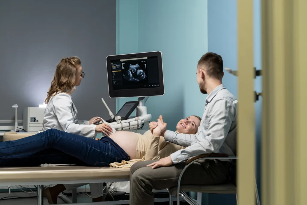
Modern prenatal care includes several advanced imaging techniques, each designed to answer different clinical questions.
High-Resolution Ultrasound
High-resolution ultrasound uses advanced ultrasound equipment to produce exceptionally detailed images of the developing baby.
It remains the foundation of fetal imaging and is often the first investigation performed when a structural abnormality is suspected.
Doppler Ultrasound
Doppler ultrasound measures blood flow within the umbilical cord, placenta, fetal brain, and other blood vessels.
It is particularly valuable for monitoring pregnancies complicated by fetal growth restriction, hypertension, pre-eclampsia, or placental insufficiency.
Fetal Echocardiography
Fetal echocardiography is a specialized ultrasound examination that evaluates the baby’s heart, including its chambers, valves, major blood vessels, and blood flow patterns.
It is recommended when congenital heart disease is suspected or when certain maternal or fetal risk factors are present (Deng et al. 2006).
3D and 4D Ultrasound
3D and 4D ultrasound provide additional anatomical information in selected pregnancies.
While three-dimensional ultrasound produces detailed still images, four-dimensional ultrasound displays those images in real time, allowing healthcare professionals to observe fetal movement.
These technologies complement, rather than replace, conventional 2D ultrasound.
Fetal MRI
Fetal magnetic resonance imaging (MRI) provides highly detailed images of the baby’s brain and other organs when ultrasound findings require further clarification.
MRI is most commonly used as a second-line imaging technique and is particularly valuable for evaluating complex neurological and thoracic abnormalities.
When Do Doctors Recommend Advanced Fetal Imaging?
Not every pregnancy requires specialised imaging.
Your healthcare provider will recommend advanced fetal imaging only when the additional information is expected to influence diagnosis, pregnancy monitoring, delivery planning, or treatment decisions.
For example, a detailed fetal echocardiogram may be recommended if there is a suspected heart defect, while fetal MRI may be requested when ultrasound cannot fully evaluate the developing brain.
Similarly, Doppler ultrasound may be performed to monitor placental function in pregnancies affected by fetal growth restriction or pre-eclampsia.
The choice of imaging technique depends on the clinical situation rather than the availability of newer technology.
By selecting the most appropriate examination for each pregnancy, healthcare professionals can improve diagnostic accuracy while avoiding unnecessary investigations.
Benefits of Advanced Fetal Imaging Techniques
Advanced fetal imaging has significantly improved prenatal care by allowing healthcare professionals to obtain more detailed information about:
the developing baby than is possible with routine ultrasound alone.
Rather than replacing standard prenatal scans, these techniques are used selectively to answer specific clinical questions and support informed decision-making throughout pregnancy.
One of the greatest benefits is earlier and more accurate diagnosis of certain fetal conditions.
When a routine ultrasound identifies an unexpected finding or provides limited visualization,
advanced imaging techniques such as high-resolution ultrasound, fetal echocardiography, Doppler ultrasound, or fetal MRI can provide additional anatomical and functional information.
This allows healthcare teams to confirm or rule out suspected abnormalities, determine their severity, and develop an appropriate management plan.
Another important advantage is improved pregnancy planning.
When a fetal abnormality is identified before birth, obstetricians, fetal medicine specialists, neonatologists, pediatric surgeons, cardiologists, and genetic counsellors can work together to plan delivery and immediate newborn care.
In some situations, knowing about a condition before birth allows delivery to be arranged at a specialized center with the appropriate facilities and expertise (Mangla et al. 2024).
High-Resolution Ultrasound
High-resolution ultrasound remains the cornerstone of advanced fetal imaging.
Modern ultrasound systems produce exceptionally detailed images that help clinicians evaluate fetal anatomy, growth, amniotic fluid volume, placental position, and multiple pregnancies.
It is usually the first advanced investigation performed when additional anatomical assessment is required because it provides real-time imaging, is widely available, and has an excellent safety profile.
Doppler Ultrasound
Unlike conventional ultrasound, Doppler ultrasound evaluates blood flow rather than anatomy alone.
It measures circulation within the umbilical cord, placenta, fetal brain, and other important blood vessels.
Doctors commonly recommend Doppler ultrasound when monitoring:
- Fetal growth restriction
- Placental insufficiency
- Pre-eclampsia
- Maternal hypertension
- Twin pregnancies with suspected complications
Assessing blood flow helps clinicians understand whether the baby is receiving adequate oxygen and nutrients.
Abnormal Doppler findings may indicate that closer monitoring or earlier delivery is required to improve pregnancy outcomes.
Fetal Echocardiography
Congenital heart disease is among the most common birth defects, making detailed assessment of the fetal heart particularly important when risk factors are present.
Fetal echocardiography is a specialised ultrasound examination that evaluates:
- Heart chambers
- Heart valves
- Major blood vessels
- Cardiac rhythm
- Blood flow through the heart
This examination may be recommended if :
there is a family history of congenital heart disease, abnormal findings on a routine anatomy scan, maternal diabetes, certain infections, or suspected chromosomal abnormalities.
Early diagnosis allows healthcare teams to prepare for appropriate monitoring during pregnancy and specialized care immediately after birth if needed.
Three-Dimensional and Four-Dimensional Ultrasound
Three-dimensional (3D) and four-dimensional (4D) ultrasound provide additional anatomical detail in selected pregnancies.
Three-dimensional ultrasound creates detailed still images that may improve visualisation of facial structures, limbs, and selected skeletal abnormalities.
Four-dimensional ultrasound builds on this technology by displaying those images in real time, allowing clinicians to observe fetal movements during the examination.
Although these technologies are widely recognized for producing lifelike images of the baby, their primary clinical role is to complement conventional ultrasound when additional anatomical information is required.
They do not replace routine prenatal imaging.
Fetal MRI
Fetal magnetic resonance imaging (MRI) is generally used when ultrasound cannot fully answer an important clinical question.
Unlike ultrasound, MRI produces highly detailed images of soft tissues and is particularly valuable for evaluating:
- The fetal brain
- Spinal abnormalities
- Chest abnormalities
- Selected abdominal conditions
MRI does not replace ultrasound. Instead, it acts as a complementary investigation that helps clarify complex findings and supports treatment planning in selected pregnancies (Gatta et al. 2021).
Limitations of Advanced Fetal Imaging
Despite remarkable technological advances, no imaging technique is perfect.
Image quality can be affected by several factors, including fetal position, gestational age, maternal body habitus, reduced amniotic fluid, and fetal movement during the examination.
In some cases, repeating the scan at a later date provides better visualisation.
Advanced imaging also cannot detect every congenital condition.
Some abnormalities develop later in pregnancy, while others may not become apparent until after birth.
For this reason, imaging findings are always interpreted together with maternal history, laboratory investigations, genetic testing when appropriate, and clinical assessment.
Are Advanced Fetal Imaging Tests Safe?
One of the most common concerns among expecting parents is whether these specialised imaging techniques are safe.
Routine ultrasound, Doppler ultrasound, fetal echocardiography, 3D ultrasound, and 4D ultrasound all use high-frequency sound waves rather than ionizing radiation.
When performed by trained healthcare professionals following recognized safety recommendations, they are considered safe during pregnancy.
Fetal MRI also does not use ionizing radiation. Instead, it uses magnetic fields and radiofrequency waves to create detailed images.
Current evidence supports its use when clinically indicated, particularly after the first trimester, and when additional diagnostic information is expected to benefit pregnancy management.
Questions to Ask Your Healthcare Provider
If advanced fetal imaging has been recommended, consider asking:
- Why is this imaging test recommended?
- What information will it provide?
- Will it change my pregnancy management?
- Are there any alternatives?
- Will I need additional testing?
- Who will explain the results?
- Should I see a fetal medicine specialist or genetic counsellor?
Understanding the purpose of the examination can reduce anxiety and help you participate confidently in decisions about your pregnancy.
Final Thoughts
Advances in prenatal imaging have transformed pregnancy care by allowing healthcare professionals to detect, monitor, and better understand many fetal conditions before birth. While these technologies provide remarkable detail, they are most effective when used for the right clinical reasons and interpreted by experienced specialists.
If your healthcare provider recommends an advanced imaging test, remember that the goal is to gather the information needed to provide the safest and most appropriate care for you and your baby. In many cases, these examinations provide reassurance and help families prepare confidently for the remainder of the pregnancy.
Conclusion
Advanced fetal imaging techniques have transformed prenatal care by allowing healthcare professionals to evaluate fetal anatomy, blood flow, heart function, and organ development with greater precision.
While most pregnancies can be monitored successfully using routine ultrasound, specialised imaging plays an important role when additional information is needed to guide diagnosis and treatment.
The most reassuring message for expecting parents is that these investigations are recommended to improve care, not to create unnecessary concern.
By combining advanced technology with experienced clinical expertise, healthcare providers can make well-informed decisions that support the best possible outcomes for both mother and baby.
Frequently Asked Questions
What is advanced fetal imaging?
Advanced fetal imaging includes specialised prenatal imaging techniques such as Doppler ultrasound, fetal echocardiography, 3D ultrasound, 4D ultrasound, and fetal MRI that provide additional information beyond routine pregnancy scans.
When is advanced fetal imaging recommended?
Your healthcare provider may recommend advanced imaging if a routine ultrasound identifies an abnormality, if your pregnancy is considered high risk, or if additional evaluation is needed.
Is fetal MRI safe during pregnancy?
Yes. Fetal MRI does not use ionising radiation and is considered safe when performed for appropriate medical reasons.
Does every pregnant woman need advanced fetal imaging?
No. Most uncomplicated pregnancies are monitored successfully using routine ultrasound without requiring additional imaging.
Can advanced fetal imaging detect birth defects?
Advanced imaging can improve the detection and assessment of many fetal abnormalities, but no single imaging test can identify every condition before birth.
Stay tuned with us for more health related topics.
Follow us on LinkedIn and Instagram for more.
More Read


Medical Disclaimer!
This article has been reviewed and written under the guidance of our Head Physiotherapist, Dr. Kruti Raj (PT, MUHS,CPT,CMPT). The information shared is intended for educational purposes only and should not be considered a substitute for personalized medical advice, diagnosis, or treatment.
Please consult us or any other qualified healthcare professional before beginning any exercise program, especially if you are experiencing pain, recovering from injury, or managing a medical condition.


