


A talus fracture is a break in the bone that connects the lower leg to the foot and helps transfer body weight through the ankle.
It often follows high-impact trauma, such as a road traffic accident, a fall from a height or a severe ankle injury.
Quick Answer: Talus Fracture
A talus fracture is a break in the ankle bone that connects the lower leg to the foot. It can cause deep ankle pain, swelling, bruising and difficulty bearing weight.
- Common causes: Falls, road traffic accidents and high-impact sports injuries.
- Diagnosis: Clinical examination and imaging, usually X-rays and sometimes CT or MRI.
- Treatment: Immobilization and restricted weight-bearing for selected stable fractures, or surgery for certain displaced or unstable injuries.
- Physiotherapy: Gradual restoration of movement, strength, balance and walking after medical clearance.
- Recovery: Often takes several months, depending on the fracture and complications.
Important: Suspected talus fractures need prompt medical assessment. Do not put weight on the injured foot unless instructed to do so.
Because the talus supports two important joints and has a vulnerable blood supply, recovery requires careful treatment and follow-up.
Physiotherapy can help restore movement, strength and walking ability, but its timing depends on fracture stability and medical clearance.
Key Takeaways
Deep ankle pain, swelling and difficulty bearing weight after trauma may indicate a fracture rather than a routine sprain.
X-rays are commonly used first, while CT or MRI may be needed to investigate more complex or occult injuries.
Follow the prescribed immobilisation and weight-bearing restrictions. Pain reduction alone does not confirm healing.
Osteonecrosis, post-traumatic arthritis and delayed healing may affect recovery in some patients.
Restore movement, strength, balance and walking gradually, according to medical clearance and functional progress.
What Is a Talus Fracture?
The talus is located between the tibia and fibula above and the heel bone, or calcaneus, below.
It forms part of the ankle and subtalar joints, allowing the foot to move up and down and adapt to uneven surfaces.
A talus fracture may involve the neck, body, head or smaller projections of the bone.
Some fractures remain aligned, while others are displaced or extend into the joint surfaces.
The talus is particularly important because much of its surface is covered with cartilage, leaving relatively limited areas for blood vessels to enter.
Damage to its blood supply can affect healing and increase the risk of complications. (Schwartz et al., 2020)
What Causes a Talus Fracture?
Most significant talus fractures occur when substantial force passes through the foot and ankle.
- Road traffic accidents: Force transmitted through the foot, particularly when the ankle is forced upwards, can fracture the talus.
- Falls from a height: Landing heavily on the feet can compress the bone between the lower leg and heel.
- Sports injuries: High-impact landings, collisions and forceful twisting may cause fractures.
- Severe ankle trauma: A fracture may accompany an ankle dislocation or other injuries to the foot and lower leg.
Less commonly, a fracture may occur with lower-impact activity when the bone is weakened.
The mechanism of injury helps clinicians determine which injuries and complications to investigate.
Symptoms of a Talus Fracture
Symptoms depend on the location and severity of the fracture.
A significant injury usually causes pain and swelling around the ankle or hindfoot.
Deep ankle pain
Pain may be felt inside the ankle or around the hindfoot.
It often worsens when standing, walking or attempting to move the foot.
Swelling and bruising
Swelling can develop rapidly, and bruising may spread around the ankle and foot over the following days.
Difficulty bearing weight
Walking may be extremely painful or impossible.
Avoid testing the injured foot by repeatedly trying to stand if a fracture is suspected.
Restricted movement
Moving the ankle upwards, downwards or from side to side may be painful.
Swelling, joint damage and immobilisation can also limit movement.
Deformity or skin injury
Displaced fractures may alter the shape of the ankle or foot.
Severe injuries can damage the skin or expose bone.
A visibly deformed ankle, an open wound, a cold or pale foot, or new numbness requires urgent medical attention.
Types of Talus Fractures
The fracture location influences treatment, complications and rehabilitation.
Talar neck fracture
The neck connects the head of the talus to its body.
Displaced fractures in this region can disrupt the bone’s blood supply and increase the risk of osteonecrosis.
Talar body fracture
The body forms a major part of the ankle joint.
Fractures may damage the joint surface and contribute to post-traumatic arthritis.
Talar head fracture
The head connects the talus to the navicular bone in the midfoot.
These fractures may affect foot mechanics and joint stability.
Lateral and posterior process fractures
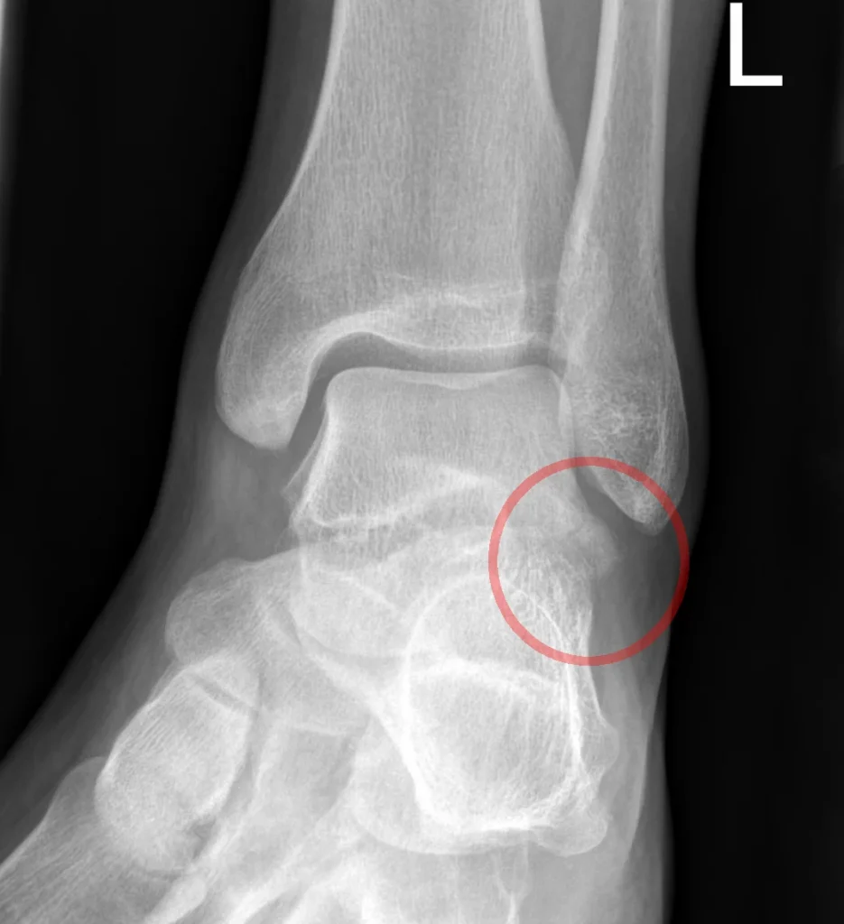
These involve smaller projections of the talus.
Some occur during sports or twisting injuries and may resemble an ankle sprain, making accurate diagnosis particularly important.
How Is a Talus Fracture Diagnosed?
Early diagnosis helps clinicians assess fracture alignment, joint involvement and the risk of complications.
Physical examination
The clinician examines swelling, bruising, skin condition, tenderness, circulation and sensation.
Associated injuries to the ankle, foot or lower leg must also be considered.
Forceful movement tests should be avoided when a fracture is suspected.
X-rays
X-rays are usually the first imaging investigation.
They can show many fractures and help assess alignment, although some small or complex injuries may not be clearly visible.
CT scans and MRI
A CT scan provides detailed images of the bone and helps identify fracture fragments, displacement and joint involvement.
It is often valuable for surgical planning.
MRI may be used selectively when an occult fracture, bone injury or associated soft-tissue problem remains suspected despite initial imaging.
Persistent clinical suspicion should not be dismissed solely because an initial X-ray appears normal. (Barnett et al., 2017)
Talus Fracture Treatment Options
Treatment depends on the fracture location, displacement, joint stability, soft-tissue condition and the person’s overall health.
Non-surgical treatment
Selected stable, nondisplaced fractures may be managed with a cast or other immobilisation, together with strict or partial restrictions on weight-bearing.
Crutches or another walking aid may be needed.
Follow-up appointments and imaging help the orthopaedic specialist monitor alignment and healing.
Surgical treatment
Displaced, unstable or joint-involving fractures often require surgery.
The surgeon may reposition the bone fragments and secure them with screws, plates or other fixation devices.
Open fractures, dislocations and injuries affecting circulation require urgent assessment.
Definitive surgery may be timed according to the condition of the surrounding soft tissues.
Important: Do not begin weight-bearing or ankle exercises simply because pain has reduced. Pain relief does not confirm that the fracture is sufficiently stable to tolerate loading.
Talus Fracture: Myth vs Fact
MYTH: If I can move my ankle, it cannot be fractured.
FACT: Some fractures still allow limited movement. Clinical assessment and appropriate imaging are needed to determine whether a fracture is present.
MYTH: A talus fracture is just a severe ankle sprain.
FACT: A fracture involves the bone and may affect the joint surface or blood supply. It needs an appropriate fracture-management plan.
MYTH: Less pain means the fracture has healed.
FACT: Symptoms can improve before the bone is ready for loading. Follow-up and clinical guidance determine when activity can progress.
MYTH: Physiotherapy should start with strengthening exercises immediately.
FACT: Rehabilitation must match the healing stage. Early priorities may include safe mobility and protection, while strengthening is introduced when appropriate.
Complications of a Talus Fracture
Some complications develop during healing, while others become apparent months or years later.
Avascular necrosis
Avascular necrosis, also called osteonecrosis, occurs when part of the bone loses its blood supply and begins to deteriorate.
The risk is particularly concerning after severe or displaced talar neck fractures.
Post-traumatic arthritis
Damage to the ankle or subtalar joint surfaces may lead to arthritis.
Symptoms can include persistent pain, stiffness and discomfort during walking.
Delayed union or nonunion
Delayed union means the fracture is taking longer than expected to heal.
Nonunion occurs when the bone does not unite adequately and may require further treatment.
Stiffness and muscle weakness
Immobilisation and restricted weight-bearing can reduce ankle mobility, calf strength, balance and walking confidence.
These problems are important targets for physiotherapy once loading and movement are permitted.
A systematic review of surgically treated talar neck and body fractures found substantial rates of complications, including osteonecrosis and arthritis.
These findings reinforce the importance of follow-up, although individual risks vary with fracture severity and treatment. (Wijers et al., 2022)
Talus Fracture Recovery Time
There is no single recovery timeline for every talus fracture.
The location of the break, displacement, surgical treatment, bone healing and complications all influence progress.
Early recovery: Protecting the fracture
During the initial weeks, treatment focuses on protecting the bone, controlling swelling and maintaining safe mobility.
Some patients must avoid putting any weight on the affected foot for an extended period.
Progressive recovery: Restoring movement
When permitted by the treating specialist, gentle ankle movement may be introduced to address stiffness.
The timing depends on fracture stability, fixation and the need to protect healing tissues.
Later recovery: Walking and daily activities
Once sufficient healing is confirmed, weight-bearing and strengthening can progress gradually.
Returning to prolonged walking, stairs, work or sport may take several months or longer, particularly after complex fractures.
Persistent pain, swelling or deteriorating function should be assessed rather than managed by simply pushing through rehabilitation.
Physiotherapy After a Talus Fracture
Physiotherapy helps restore function after a talus fracture, but the programme must match the stage of healing.
Exercises appropriate after a stable fracture has healed may be unsafe during early fracture protection.
1. Safe mobility and swelling management
Early rehabilitation may focus on using crutches correctly, transferring safely and maintaining mobility without loading the injured foot beyond the prescribed limit.
Elevation may help manage swelling when appropriate.
Exercises for unaffected joints and body regions can help maintain general mobility during immobilisation.
2. Gentle ankle range-of-motion exercises
When the orthopaedic team permits movement, controlled ankle exercises can help address stiffness and improve mobility.
Depending on the fracture and treatment, these may include gentle ankle movements within a comfortable range.
Forceful stretching and unsupervised movements that stress the healing bone should be avoided.
3. Gradual strengthening
After sufficient healing and clearance for progressive loading, physiotherapy may introduce exercises for the calf and muscles that support the ankle and foot.
Resistance, repetitions and exercise difficulty should increase gradually, guided by the treatment plan and the person’s response.
4. Balance and walking retraining
After immobilisation, people may feel unsteady or develop an altered walking pattern.
Once weight-bearing is permitted, rehabilitation may include supported standing, gait retraining and progressively challenging balance exercises.
These activities help rebuild coordination and confidence during daily movement.
5. Return to work, running and sport
Returning to demanding activities requires more than the ability to walk without a limp.
The person may also need adequate strength, joint mobility, balance and tolerance for repeated loading.
A physiotherapist can help develop a graded return-to-activity plan in coordination with the orthopaedic team.
Running and jumping should wait until healing and functional readiness have been assessed.
What should be avoided during rehabilitation?
Until the treating specialist gives clearance, avoid:
- Putting weight through the foot against medical instructions.
- Starting calf raises, hopping or impact exercises prematurely.
- Forcing ankle movement to overcome stiffness.
- Progressing exercises despite increasing pain or swelling.
- Treating the injury as an ordinary ankle sprain without confirming fracture stability.
Physiotherapy supports recovery, but it cannot replace fracture fixation, immobilisation or medical monitoring when these are required.
When Should You Seek Medical Help?
Seek prompt medical assessment after a significant ankle injury, particularly if you cannot bear weight or have severe swelling and pain.
Go to an emergency department immediately if the foot or ankle is visibly deformed, bone is exposed, the foot becomes cold or pale, or you develop new numbness or rapidly worsening pain.
After treatment, contact your clinical team if pain or swelling increases unexpectedly, a cast feels excessively tight, a wound becomes concerning or your symptoms change significantly.
Lesser-Known Facts About Talus Fractures
A small fracture can resemble an ankle sprain
Fractures of the lateral or posterior talar processes may produce symptoms similar to a sprain.
Persistent pain after a twisting injury deserves reassessment, especially when the initial diagnosis is uncertain.
Bone healing and joint recovery are different
A fracture may unite while cartilage damage, stiffness or post-traumatic arthritis continues to affect function.
Follow-up should consider both bone healing and the person’s symptoms and mobility.
Osteonecrosis may require long-term monitoring
A person may need follow-up imaging and clinical assessment after the fracture has healed.
The risk depends on the fracture pattern and severity, and not every patient develops osteonecrosis.
Rehabilitation is individual, not calendar-based
Progressing to weight-bearing, strengthening or sport should depend on clinical findings and the treating team’s instructions, rather than a fixed number of weeks.
Final Thoughts: Recovering After a Talus Fracture
A talus fracture needs careful management because it can affect bone healing, ankle movement and long-term joint health. Prompt assessment and appropriate follow-up help clinicians identify complications and guide recovery.
Physiotherapy can help rebuild movement, strength, balance and walking ability once the fracture is ready for rehabilitation. The right programme is gradual and tailored to the injury, rather than based on a fixed recovery schedule.
Remember: Protect the healing bone, follow your weight-bearing instructions and progress activity only when your treating team says it is safe.
Conclusion
A talus fracture can affect ankle movement, walking and long-term joint health.
Early diagnosis, appropriate fracture management and follow-up are essential because some injuries can compromise the talus’s blood supply or damage its joint surfaces.
Physiotherapy helps address stiffness, weakness, balance problems and walking difficulties once the fracture can safely tolerate rehabilitation.
A gradual, medically guided programme can support a return to daily activities while accounting for individual healing and possible complications.
Frequently Asked Questions
1. How serious is a talus fracture?
A talus fracture can be serious because it may affect the ankle joint, bone blood supply and ability to walk. Severity depends on the fracture location, displacement and associated injuries.
2. How long does a talus fracture take to heal?
Healing and functional recovery vary. Some fractures need several months of treatment and rehabilitation, while complex injuries or complications can extend recovery. Your orthopaedic specialist should guide your timeline.
3. Can a talus fracture heal without surgery?
Some stable, nondisplaced fractures can be treated without surgery using immobilisation and prescribed weight-bearing restrictions. Displaced or unstable fractures may require an operation.
4. Can physiotherapy help after a talus fracture?
Yes. Once medically cleared, physiotherapy can help restore ankle movement, muscle strength, balance, walking mechanics and tolerance for daily activities. It does not replace fracture protection or medical treatment.
5. When can I walk after a talus fracture?
This depends on the fracture pattern, treatment and evidence of healing. Some patients must avoid weight-bearing for an extended period. Do not start walking without the clearance specified by your treating team.
6. Can a talus fracture cause arthritis later?
Yes. Damage to the ankle or subtalar joint can contribute to post-traumatic arthritis. The risk varies with the injury and the degree of joint-surface damage.
Stay tuned with us for more health related topics.
Follow us on LinkedIn and Instagram for more.
More Read


Medical Disclaimer
This article is intended for general educational purposes only and does not replace professional medical advice, diagnosis or treatment. A suspected talus fracture requires prompt medical assessment. Do not begin weight-bearing, exercises or rehabilitation without following the instructions of your orthopedic specialist and physiotherapist. Seek urgent medical care for severe pain, visible deformity, an open wound, new numbness or a cold or pale foot.


