


LONDON, UK: Five artificial intelligence (AI) tools have been recommended in draft guidance from the National Institute for Health and Care Excellence (NICE) to help detect prostate cancer on MRI scans in England.
The technologies are intended to support radiologists in identifying suspicious areas and improving diagnostic consistency.
NICE has proposed their use over a two-year period while further evidence is collected on their effectiveness. The evaluation will examine diagnostic accuracy, patient outcomes, clinical workflows and performance across different patient groups.
AI Tools Support Prostate Cancer Detection
Prostate MRI plays an important role in investigating suspected prostate cancer. Radiologists examine these scans to identify suspicious lesions and assess whether further investigations, including a biopsy, may be necessary.
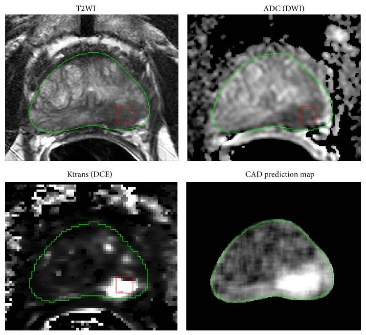
AI-assisted software can analyse prostate MRI images and highlight areas where cancer may be present. This helps radiologists focus on suspicious findings and supports the interpretation and reporting process.
According to Pulse Today’s report on the NICE recommendation, the technologies could act as a second pair of eyes for radiologists. They may help improve reporting consistency and identify scans that require priority review.
Five AI tools received a positive recommendation in the draft guidance, while two additional tools were not recommended because more research was needed before routine use could be supported.
The recommendation is not a claim that AI can diagnose prostate cancer independently. Radiologists will continue to review the original MRI images and the software’s findings before making reporting decisions.
How AI Analyses Prostate MRI Scans
AI-assisted prostate MRI software processes medical images to identify patterns and abnormalities that may indicate cancer. Depending on the technology, it can highlight suspicious regions, classify findings and provide information that supports the radiologist’s assessment.
The software is used after MRI acquisition, alongside the existing diagnostic process. It does not remove the need for specialist interpretation or replace the clinical assessment of the patient.
Radiologists must consider AI-generated findings alongside the patient’s medical history, prostate-specific antigen (PSA) results and other relevant investigations.
A suspicious MRI finding does not automatically confirm cancer, and the absence of an AI alert does not independently rule it out.
The purpose of these tools is to support clinical judgement and potentially make MRI reporting more accurate and consistent.
Potential Benefits for Patients
Improved Detection Accuracy
One potential benefit is better identification of clinically significant prostate cancer. This term refers to cancer that is more likely to affect a person’s health and may require treatment.
By highlighting suspicious lesions, AI may help radiologists identify findings that need closer examination. More consistent interpretation could support clinical decision-making and reduce differences in reporting between healthcare professionals.
However, the extent of any improvement must be established through further evaluation in routine clinical settings.
Fewer Unnecessary Procedures
Not every suspicious prostate MRI finding represents clinically significant cancer. Some findings may be benign, while some cancers may grow slowly and never cause significant health problems.
If AI-assisted reporting improves the assessment of suspicious lesions, it could help clinicians make better-informed decisions about further investigations. This may reduce unnecessary biopsies and help avoid treatment for prostate cancers that are unlikely to cause harm.
These outcomes remain potential benefits rather than established results of the five recommended tools. Decisions about biopsy and treatment must continue to reflect the patient’s individual circumstances and clinical assessment.
More Efficient MRI Reporting
Demand for prostate MRI scans has increased in recent years, adding pressure to radiology services. AI may help radiologists review scans more efficiently by directing attention towards areas that warrant further examination.
The technologies could also help clinical teams identify scans that need priority review. This may support more efficient use of available diagnostic capacity.
However, actual improvements in reporting times and waiting lists will depend on how the software performs and how hospitals incorporate it into their existing workflows.
Why NICE Requires More Evidence
NICE has proposed a two-year period of evidence collection to assess how the five recommended tools perform in routine NHS practice. Two additional technologies were not recommended because their effectiveness required further research.
The evaluation will focus on several important areas.
- Diagnostic accuracy: How reliably the software identifies clinically significant cancer and distinguishes it from less significant findings.
- Patient outcomes: Whether AI-assisted reporting leads to better-informed clinical decisions and improved patient care.
- Clinical workflows: How the technology affects reporting time, radiologists’ workloads and the efficiency of diagnostic services.
- Cost-effectiveness: Whether the clinical and operational benefits justify the cost of implementing and maintaining the software.
- Performance across patient groups: Whether the technology delivers consistent results for different groups of patients and across different NHS settings.
These assessments are important because AI performance can vary with image quality, MRI protocols and patient characteristics. Evidence from one clinical setting may not fully predict how a system performs in another.
Radiologists Remain Responsible for Diagnosis
NICE has emphasized that AI will support, not replace, radiologists. Healthcare professionals will continue to review the scan images, assess the software’s findings and make the final reporting decisions.
Dr Adrian Hayter, GP and chief medical officer at NICE, said the tools could help radiologists identify clinically significant prostate cancer more accurately and consistently while supporting NHS services facing growing demand.
Dr Anastasia Chalkidou, program director for HealthTech at NICE, highlighted the potential for the technology to improve MRI reporting and help identify scans that may need priority review.
She also emphasized the need to gather further evidence on performance in routine practice and across different patient groups.
Professor Peter Johnson, NHS national clinical director for cancer, welcomed the opportunity to evaluate these technologies. He referred to the potential of AI innovations, including those used in lung cancer diagnosis, to support faster cancer detection.
These developments highlight the importance of combining technological assistance with qualified clinical oversight. AI findings must be interpreted in context, and clinical decisions must remain based on the complete patient assessment.
Prostate Cancer Diagnosis in England
More than 58,000 new cases of prostate cancer were diagnosed in England in 2023, according to the source report. This substantial diagnostic demand reinforces the need for reliable investigation pathways and efficient use of specialist services.
PSA testing and MRI serve different roles in assessing prostate cancer risk. A raised PSA level does not necessarily mean cancer is present, while a result within the expected range does not exclude every case.
MRI can help clinicians investigate suspected prostate cancer and guide decisions about whether a biopsy is appropriate. When a biopsy is performed, tissue samples are examined to establish whether cancer is present and assess its characteristics.
In August, NHS England issued updated advice to GPs on when to offer PSA testing. The National Screening Committee had advised against routine population-wide NHS screening for prostate cancer, recommending a targeted approach for certain higher-risk men.
The targeted recommendation involved PSA testing every two years for men aged 45 to 61 who have a pathogenic BRCA2 variant and a family history of breast, ovarian, pancreatic or prostate cancer.
This applies to a specific higher-risk group rather than the general male population.
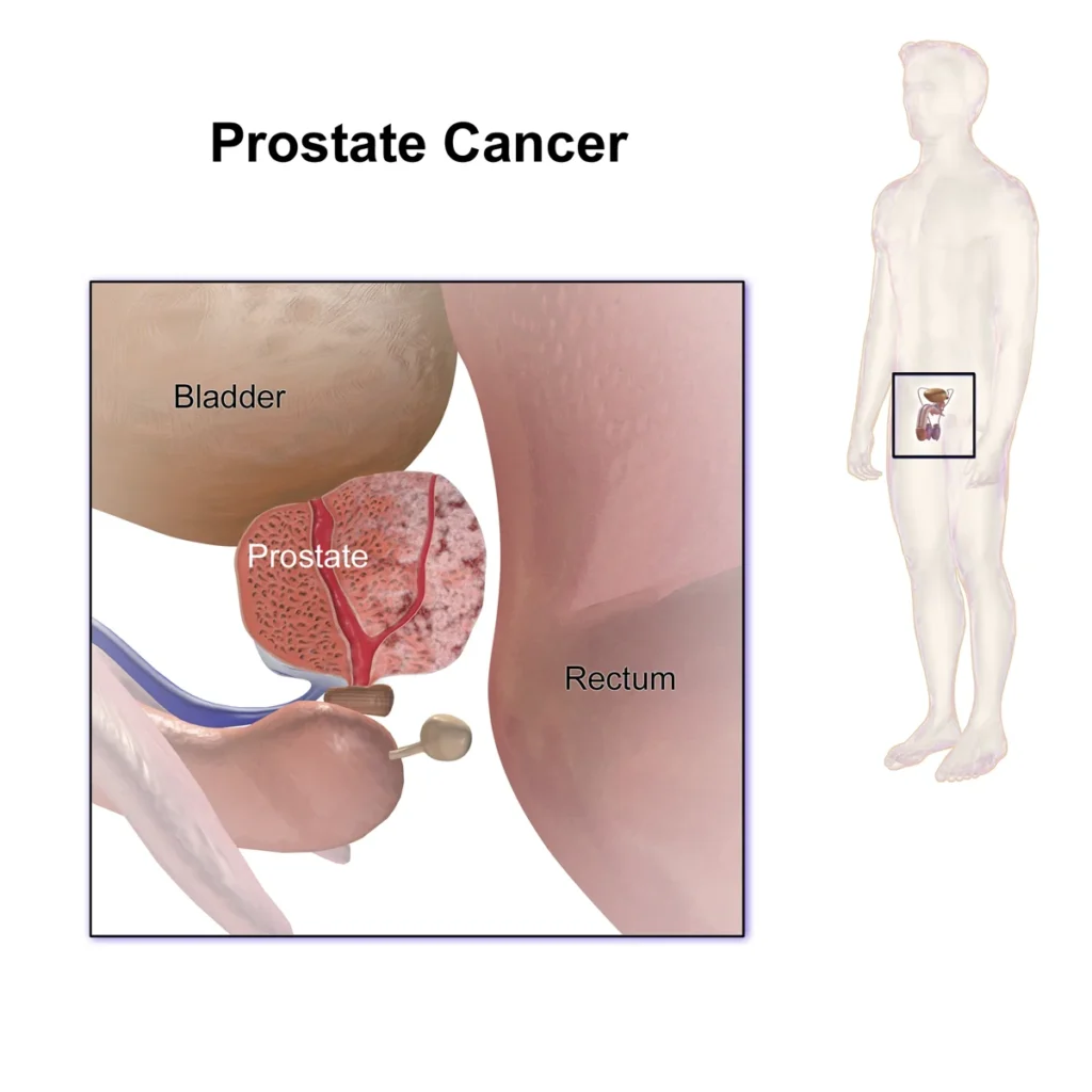
The AI recommendation concerns the assessment of prostate MRI scans for suspected cancer. It should not be confused with a population-wide screening program or a replacement for existing diagnostic pathways.
What the Recommendation Means
The draft NICE recommendation represents a step towards evaluating AI-assisted prostate MRI reporting within routine NHS services.
The technology may improve diagnostic consistency, help identify clinically significant cancer and support more efficient use of radiology resources.
However, its long-term value will depend on the evidence collected during the proposed two-year period. Diagnostic accuracy, patient outcomes, cost-effectiveness and performance across different groups will all influence future decisions about routine use.
For patients, AI may eventually contribute to a more efficient diagnostic pathway. For radiologists, it offers an additional tool to support the interpretation of complex medical images without removing professional responsibility.
Conclusion
Five AI tools have been recommended in draft NICE guidance to support prostate cancer detection on MRI scans, while two additional tools require further research.
The technologies could help radiologists identify clinically significant cancer more consistently, support prioritization of scans and reduce unnecessary biopsies and treatment.
Further evidence is needed to establish whether these potential benefits translate into better patient outcomes and more efficient NHS services.
Until that evidence is available, AI should remain a support tool used alongside specialist radiological interpretation and comprehensive clinical assessment.