


Can accessory navicular cause ankle pain?
Yes, it can cause pain around the inner foot and medial ankle, particularly when the accessory navicular bone irritates surrounding tissues or affects the function of the posterior tibial tendon.
If you have pain on the inside of your foot or ankle, especially around the inner arch, and you have noticed a hard or prominent bump in that area, an accessory navicular could be part of the explanation.
The interesting part is that an accessory navicular is not necessarily a disease.
It is an extra bone or ossicle near the navicular bone, and many people have one without ever knowing it.
The problem begins when that structure becomes irritated, overloaded, or mechanically involved with the posterior tibial tendon and the arch of the foot.
So, can an accessory navicular cause foot and ankle pain?
Yes, it can.
But the presence of an accessory navicular on an X-ray does not automatically mean it is the source of your pain.
Quick Answer
Yes. Accessory navicular syndrome can cause pain on the inner side of the foot and may sometimes be perceived as inner ankle pain. However, simply having an accessory navicular on an X-ray does not mean it is the source of symptoms. Pain is more likely when the accessory navicular becomes irritated through repetitive loading, footwear pressure, posterior tibial tendon stress or associated flatfoot mechanics. Treatment usually begins with activity modification, appropriate footwear, orthotic support when indicated and progressive physiotherapy rather than immediate surgery.
That distinction is extremely important in physiotherapy.
I often see people become worried after an imaging report says “accessory navicular,” even though the finding may simply be an incidental anatomical variation.
A 2024 meta-analysis involving 39 studies and more than 36,000 feet estimated that accessory navicular occurs in about 12.6% of feet, with approximately half of affected people having it on both sides. Most are asymptomatic. [SpringerLink]
More recently, an adult MRI study published in 2026 found accessory navicular in 24.4% of 1,988 ankle MRI examinations, again showing how frequently this anatomical variation can appear on imaging.
Type II was the dominant subtype, and bone marrow edema was particularly associated with Type II accessory navicular. [MDPI]
The real question, therefore, is not simply, “Do I have an accessory navicular?”
It is:
“Is my accessory navicular actually causing my pain?”
Key Takeaways
- An accessory navicular is a developmental extra bone near the inner side of the foot.
- Most accessory navicular bones are painless and do not require treatment.
- Type II accessory navicular is particularly relevant in symptomatic cases.
- Pain usually occurs around the medial arch or inner side of the foot and may sometimes feel like inner ankle pain.
- Posterior tibial tendon loading, flexible flatfoot, footwear pressure and sudden increases in activity can contribute to symptoms.
- An X-ray showing an accessory navicular does not automatically prove that it is causing the pain.
- MRI can help identify bone marrow edema, soft-tissue irritation and associated tendon problems when clinically indicated.
- Physiotherapy may include activity modification, footwear changes, orthotic support, mobility work and progressive foot and posterior tibial strengthening.
- Surgery is generally considered when appropriately managed conservative treatment fails or when significant associated deformity or tendon problems are present.
What exactly is an accessory navicular?
The navicular is one of the important bones in the middle of your foot.
It sits between the talus and the cuneiform bones and forms part of the medial longitudinal arch.
An accessory navicular is an additional piece of bone or cartilage located on the inner side of the navicular.
It is developmental rather than something that usually appears because you injured your foot.
You may also hear it called:
- Accessory navicular bone
- Os naviculare
- Os tibiale externum
- Accessory tarsal navicular
- Navicular syndrome, in some clinical contexts
The accessory navicular is particularly interesting because of its relationship with the tibialis posterior tendon.
The tibialis posterior muscle and tendon help support the medial arch, control foot movement and assist with inversion and plantarflexion.
In some people, the tendon attaches partly or substantially around the accessory navicular.
That means the accessory bone is not simply an isolated “extra bone.”
It can become part of a mechanical system involving the tendon, arch and surrounding soft tissues.
Research reviews describe this relationship as one reason symptomatic accessory navicular can be associated with posterior tibial tendon dysfunction and flexible flatfoot. [World Journal Of Clinical Cases]
Why does an accessory navicular become painful?
There is no single mechanism.
Several factors can combine to make an otherwise harmless anatomical variation symptomatic.
Repetitive stress at the synchondrosis
Type II accessory naviculars are particularly important because they are connected to the main navicular through a cartilage-like junction called a synchondrosis.
This junction can experience repeated tension, compression and shear.
If the area is repeatedly loaded during running, jumping, sports or prolonged walking, the synchondrosis can become painful.
Older biomechanical research demonstrated that Type II accessory naviculars can experience substantial mechanical forces at this junction, providing a plausible explanation for painful symptoms. [Sella & Lawson, 1987]
This also explains why someone can have an accessory navicular for years without symptoms and then suddenly develop pain after increasing running mileage, changing sports, gaining training volume or wearing different footwear.
Irritation from footwear
This is one of the simplest but most overlooked causes.
The accessory navicular sits on the medial side of the foot, exactly where certain shoes can press against it.
Tight shoes, narrow sports shoes, rigid footwear or footwear with a firm medial edge can repeatedly rub against the prominence.
You may notice:
- Redness over the bump
- Tenderness when wearing shoes
- Pain that improves after removing footwear
- Pain after long periods of walking
- A feeling that one particular shoe “hits the bone”
In these cases, changing footwear can sometimes make a surprisingly large difference.
Increased demand on the posterior tibial tendon
If the tibialis posterior tendon has to work harder to support the arch, the accessory navicular region can become more mechanically stressed.
This may occur with:
- Flexible flat feet
- Sudden increases in running
- Repetitive jumping
- Long-distance walking
- Poorly tolerated footwear
- Reduced calf or ankle mobility
- Weakness of the foot and ankle musculature
- Changes in body weight or activity level
However, it is important not to blame flat feet automatically.
A person can have flat feet and an accessory navicular without pain.
The important issue is whether the combination is creating excessive mechanical demand and symptoms.
The three types of accessory navicular
Doctors commonly classify accessory navicular bones into three broad types.
Type I
Type I is generally a small, separate ossicle located within or near the posterior tibial tendon.
It is often incidental and may never cause symptoms.
Type II
Type II is usually the most clinically important type.
It is larger and connected to the navicular through a synchondrosis rather than being completely fused.
This area can become painful because the connection is subjected to mechanical forces.
Type II is also frequently reported in symptomatic patients.
A 2019 study of 169 pediatric patients with symptomatic accessory navicular found Type II accounted for approximately 73% of symptomatic accessory naviculae in that cohort. [Wynn et al., 2019]
Type III
Type III represents a prominent or enlarged medial portion of the navicular, often described as a fused accessory navicular or cornuate navicular.
Because the prominence is incorporated into the navicular, symptoms may be more related to local pressure, altered biomechanics and tendon loading rather than movement across a separate synchondrosis.
Interestingly, research suggests Type II morphology may sometimes progress toward a Type III appearance as skeletal maturity develops. [Journal of Children’s Orthopaedics]
What does accessory navicular pain actually feel like?
The location of the pain is one of the biggest clues.
Typical symptoms include pain or tenderness along the inner side of the midfoot, particularly around the bony prominence.
Some people describe:
- A dull ache in the medial arch
- Sharp pain after running
- Tenderness when shoes press against the area
- Pain while walking barefoot
- Pain after prolonged standing
- Pain during sports
- Pain when pushing off the foot
- Swelling around the prominence
- A visibly prominent bump
- Pain extending toward the inner ankle
The pain does not always stay directly over the accessory bone.
This is where the ankle-foot connection becomes clinically important.
Because the posterior tibial tendon travels behind the inner ankle and continues toward the navicular, irritation around its insertion can produce symptoms that patients interpret as ankle pain.
So yes, accessory navicular syndrome can cause what feels like inner ankle pain.
But if your pain is directly behind the ankle bone, around the Achilles tendon, on the outer ankle or deep inside the ankle joint, another diagnosis may be more likely.
A simple clue: the “shoe test”
One useful clinical clue is surprisingly simple.
Put on the shoe that normally aggravates your symptoms.
Then compare it with a softer, wider shoe that does not press against the medial prominence.
If symptoms appear consistently with direct pressure from the shoe and settle when that pressure is removed, footwear irritation may be contributing.
This does not prove accessory navicular syndrome, but it provides useful information during a clinical assessment.
Why an X-ray finding does not prove the accessory navicular is causing your pain
This is one of the most important points in this article.
Suppose an X-ray says:
“Accessory navicular noted.”
That does not automatically mean:
“Accessory navicular syndrome is the cause of the patient’s ankle pain.”
Accessory navicular is common in people without symptoms.
A 2024 meta-analysis found a pooled prevalence of 17.5% at the patient level, demonstrating that this is a relatively common anatomical variation. [Springer Nature]
Similarly, a study of patients with chronic foot pain found accessory navicular in approximately one-fifth of patients, but only a smaller proportion had pain specifically attributable to the accessory navicular. [Wiley]
This is why the physical examination matters.
Your pain location, tenderness, foot posture, tendon strength, gait, footwear, activity history and imaging findings should all tell the same story before the accessory navicular is considered the main pain generator.
How is accessory navicular syndrome diagnosed?
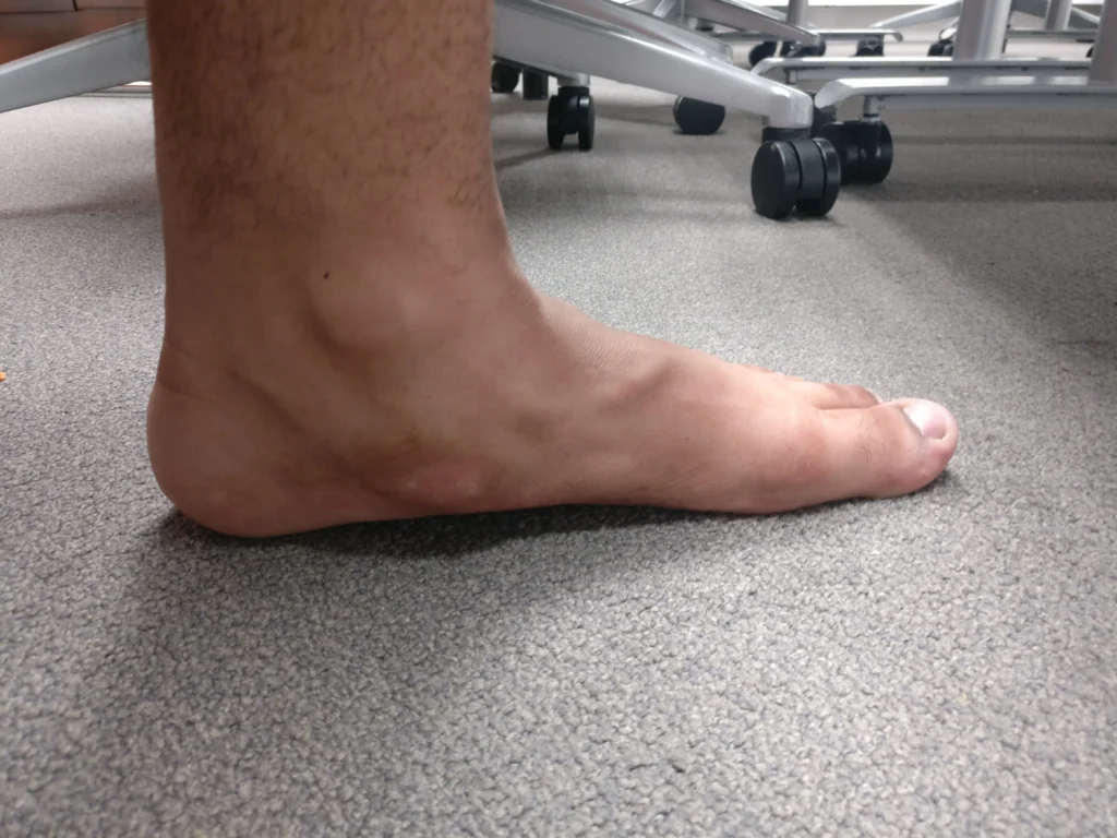
Diagnosis usually begins with a detailed history and physical examination.
Your clinician may assess:
- Exact location of tenderness
- Foot arch height
- Hindfoot alignment
- Single-leg balance
- Walking pattern
- Calf flexibility
- Ankle range of motion
- Tibialis posterior strength
- Ability to perform a single-leg heel raise
- Foot and ankle loading
- Shoe pressure
- Pain during resisted inversion and plantarflexion
Resisted activation of the posterior tibial tendon can reproduce symptoms in some patients.
A 2008 review of pediatric foot and ankle pain specifically notes that resisted posterior tibialis testing can cause or reproduce pain in symptomatic accessory navicular cases. [ Pediatric Rheumatology]
X-ray
Plain radiographs are usually the starting point.
They can demonstrate the accessory bone and help classify its type.
Weight-bearing views can also provide information about the foot’s overall alignment and whether a flexible or more significant flatfoot pattern is present.
MRI
MRI is particularly useful when the diagnosis is uncertain or symptoms are significant.
It can demonstrate:
- Bone marrow edema
- Soft-tissue inflammation
- Tibialis posterior tendon abnormalities
- Synchondrosis irritation
- Other causes of medial foot pain
Importantly, research has shown that simply seeing an accessory navicular on an X-ray may be insufficient to establish that it is painful, whereas MRI can provide evidence of bone marrow and soft-tissue edema. [Korean journal of Radiology]
A 2026 MRI study found bone marrow edema in approximately 21% of Type I and Type II accessory navicular cases assessed for edema, with Type II morphology showing a particularly strong association. [MDPI]
That makes MRI useful, but even MRI findings still need clinical correlation.
Can accessory navicular cause flat feet?
It can be associated with flat feet, but the relationship is more complicated than “accessory bone equals flat foot.”
The posterior tibial tendon is one of the structures that helps support the medial arch.
If its insertion is altered by an accessory navicular, the mechanical behavior of the tendon and arch may change.
This is particularly relevant in children and adolescents, although symptomatic cases can occur in adults too.
The combination of:
accessory navicular + posterior tibial tendon dysfunction + flexible flatfoot
can create a cycle in which the foot becomes less efficient at controlling pronation and the medial structures experience greater loading.
Recent systematic-review evidence also suggests that when symptomatic accessory navicular occurs alongside significant pes planus, correcting the broader foot alignment may influence outcomes rather than treating the accessory bone in isolation. [Sage Journals]
Physiotherapy treatment: what actually helps?
The goal of physiotherapy is not to “remove” the accessory bone.
Instead, the goal is to reduce unnecessary stress around it and improve the foot and ankle’s ability to tolerate load.
Treatment should be individualized.
1. Reduce the irritating load first
If every running session aggravates the medial foot, repeatedly pushing through the pain is usually not a rehabilitation strategy.
Temporarily reduce:
- Running
- Jumping
- Sprinting
- Long walks
- Hill training
- High-volume sports
This does not necessarily mean complete rest.
The aim is relative unloading, followed by gradual reloading.
2. Fix the shoe before blaming the foot
A wide toe box is not enough if the shoe still presses directly against the medial prominence.
Look for footwear with:
- Adequate width
- A softer medial upper
- Enough room around the arch
- Appropriate cushioning
- Stable construction when needed
Do not deliberately wear extremely soft, unsupportive shoes simply because they feel comfortable initially. Some people need more structured support.
3. Consider an orthosis when appropriate
Arch-supporting orthoses can reduce mechanical stress in selected patients.
A 2024 study of children with symptomatic accessory navicular combined with flexible flatfoot found meaningful improvements in pain and foot-related measures after custom foot orthosis treatment over four years. [Indian Journal of Orthopaedics]
A 2025 randomized study in professional athletes with accessory navicular syndrome also reported improvements in pain, arch parameters and plantar pressure distribution with customized orthoses compared with conventional insoles. [BMC]
However, an orthosis should not simply be selected because an X-ray shows an accessory navicular.
It should be chosen according to your foot posture, symptoms, footwear and loading pattern.
4. Strengthen the posterior tibial system
The posterior tibial tendon deserves attention, particularly if weakness or poor endurance is present.
A physiotherapist may prescribe progressive exercises such as:
- Resisted inversion
- Heel raises
- Single-leg heel raises
- Controlled calf raises with appropriate foot alignment
- Foot intrinsic strengthening
- Balance exercises
- Progressive functional loading
The progression matters more than the individual exercise.
If you can perform 20 easy heel raises without symptoms, doing the same 20 forever is unlikely to create meaningful adaptation.
The load should gradually increase according to symptoms and function.
Research on early posterior tibial tendon dysfunction has shown benefits from orthoses, stretching and progressive resistance exercise, with eccentric strengthening producing particularly favorable improvements in one randomized controlled trial. [Oxford Academics]
5. Do not forget the calf
A stiff calf can alter how the foot moves during walking, squatting and running.
If ankle dorsiflexion is restricted, the foot may compensate through other segments.
That does not mean every person with accessory navicular syndrome needs aggressive calf stretching.
Instead, a physiotherapist should determine whether calf restriction is actually contributing to your movement pattern.
6. Retrain the whole movement chain
This is an area where rehabilitation can become much more sophisticated.
The foot does not work independently.
Hip strength, knee control, pelvic stability, running technique and single-leg control can influence how load reaches the foot.
For a runner, for example, simply strengthening the foot may not be enough if training volume has increased from 10 km per week to 40 km per week in a short period.
The rehabilitation plan should therefore address capacity versus demand.
A useful physiotherapy rule: pain is information, not always damage
A common mistake is interpreting every pain increase as evidence that the bone is being damaged.
Pain can reflect irritation and sensitivity around the synchondrosis, tendon or soft tissues.
At the same time, persistent or worsening pain should not simply be ignored.
A useful rehabilitation approach is to monitor:
Pain during activity + pain later that day + next-morning response + functional ability.
If your symptoms repeatedly become worse the following morning after an exercise session, the load may be progressing too quickly.
This is more useful than judging rehabilitation solely by how the foot feels during the exercise.
Things you should avoid
Do not aggressively massage the bony prominence
Deep pressure directly over a painful accessory navicular can irritate an already sensitive area.
Massage may be useful for surrounding soft tissues in selected cases, but repeatedly pressing directly on the painful prominence is not a treatment by itself.
Do not force painful arch exercises
Foot strengthening should create an appropriate training stimulus, not severe localized pain.
If an exercise consistently reproduces sharp pain over the accessory navicular, discuss modification with your physiotherapist.
Do not buy the most expensive orthotic automatically
More expensive does not necessarily mean more appropriate.
The best orthosis is the one that improves your symptoms and function while fitting comfortably into the footwear you actually use.
Do not return to running simply because walking no longer hurts
Running creates substantially different loading demands.
A sensible return may progress through:
walking → brisk walking → low-impact exercise → short jog intervals → continuous running → increased volume → speed and hills.
The exact progression depends on symptoms and capacity.
When should you see an orthopaedic specialist?
You should consider medical assessment if:
- Pain persists despite several weeks of appropriate load modification
- The medial foot is significantly swollen
- You cannot participate in normal activities
- You cannot perform a controlled heel raise
- Symptoms repeatedly return despite rehabilitation
- There was a significant injury
- Pain is present at rest or increasingly at night
- There is marked redness, warmth or unexplained swelling
- You have significant flatfoot progression
- MRI shows substantial bone marrow edema or tendon pathology
Persistent symptoms do not automatically mean you need surgery.
They mean the diagnosis and treatment strategy deserve reassessment.
Does accessory navicular syndrome require surgery?
Not necessarily.
Conservative treatment is generally attempted first, particularly in children and adolescents.
However, the evidence is important because “just rest” is not always enough.
In a 2019 study of 169 pediatric patients with symptomatic accessory navicular, 28% achieved complete pain relief, 41% achieved partial relief without surgery, and approximately 30% eventually required surgery.
Complete pain relief in the successful group took an average of eight months of nonoperative treatment.
A 2024 population-based case-cohort study found that approximately 28.8% of the subcohort eventually underwent surgery after failure of nonoperative management.
Factors associated with a higher risk of failure included older age, activity limitation, higher BMI and MRI bone marrow edema. [Research Gate]
These numbers are useful for counselling patients, but they should not be interpreted as an individual prediction.
What does surgery involve?
Surgical options vary according to anatomy and the presence of flatfoot or posterior tibial tendon problems.
Procedures may include:
- Simple excision of the accessory navicular
- Kidner procedure
- Modified Kidner procedure
- Procedures addressing associated flatfoot deformity
A 2023 systematic review found generally favorable outcomes following surgical treatment, although much of the available evidence consists of retrospective studies and treatment approaches vary. [Cureus]
More recent evidence also suggests that patients with substantial flatfoot may require a more individualized approach rather than simply removing the accessory bone. [Journal of Foot and Ankle Research]
Surgery therefore should not be decided purely by the size of the accessory navicular.
A lesser-known point: the other foot may have one too
If an accessory navicular is found on one side, the other foot may also have one.
The 2024 meta-analysis estimated bilateral occurrence in approximately 50% of affected patients.
A 2026 adult MRI study reported bilateral accessory navicular in 88.4% of the subset in whom both sides were available for MRI assessment. [PubMed]
This does not mean both feet will hurt.
It simply demonstrates that the anatomical variation itself may be bilateral while symptoms remain one-sided.
Another overlooked clue: your symptoms may begin after a change, not an injury
Patients sometimes say:
“I never injured my foot. Why did this suddenly start hurting?”
That is entirely possible.
Accessory navicular syndrome can become symptomatic after a change in load rather than one dramatic injury.
For example:
You start running again after six months.
You increase your weekly mileage.
You join a dance class.
You start playing badminton every weekend.
You buy a stiffer pair of sports shoes.
You spend several days walking much more than usual.
The bone did not necessarily “appear.”
The mechanical environment changed.
This is one reason rehabilitation should investigate what changed in the weeks before symptoms started.
My physiotherapy approach to accessory navicular pain
If I were assessing a patient with suspected accessory navicular syndrome, I would not stop after confirming the bone on an X-ray.
I would ask five bigger questions:
Where exactly is the pain?
What activity triggers it?
What is happening to the medial arch during loading?
How well is the posterior tibial tendon functioning?
What changed before the pain began?
Then I would build treatment around the answers.
For one person, changing footwear may be the biggest intervention.
For another, the priority may be temporary activity modification and immobilization.
For a runner, training-load management may matter most.
For someone with flexible flatfoot and tendon weakness, orthotic support plus progressive strengthening may be more appropriate.
This is why there is no single “best exercise for accessory navicular syndrome.”
Quick prevention strategy
You cannot prevent having an accessory navicular because it is a developmental anatomical variation.
You can, however, reduce the chance of repeatedly irritating it.
Try to:
- Increase sports volume gradually
- Avoid sudden jumps in running mileage
- Wear footwear that does not press directly on the prominence
- Replace excessively worn-out shoes
- Maintain calf and ankle mobility when restricted
- Build posterior tibial and foot strength progressively
- Address persistent flatfoot mechanics when clinically relevant
- Give recurring pain proper attention instead of repeatedly pushing through it
- Return to sport gradually after a flare
One particularly useful habit is to pay attention to next-day symptoms rather than only symptoms during exercise.
If today’s workout repeatedly produces worse pain tomorrow morning, your current training dose may be too high.
Final word
Accessory navicular syndrome is a good example of why an imaging finding should never be interpreted in isolation.
You can have an accessory navicular and completely normal feet.
You can also have an accessory navicular that becomes the source of significant medial foot pain, particularly when the synchondrosis, posterior tibial tendon, footwear pressure or foot mechanics become overloaded.
The most important distinction is between having an accessory navicular and having symptomatic accessory navicular syndrome.
If your pain is located around the inner arch or inner ankle, especially if you have a prominent bump, pain with footwear, activity-related symptoms or signs of posterior tibial tendon dysfunction, an accessory navicular deserves consideration.
The encouraging part is that treatment does not automatically mean surgery.
Load modification, appropriate footwear, orthotic support when indicated, progressive strengthening and careful rehabilitation can be valuable.
At the same time, persistent symptoms deserve proper reassessment rather than endless trial-and-error exercises.
The goal is not simply to make the X-ray look different.
The goal is to make the foot strong enough, comfortable enough and mechanically efficient enough to handle the life you want to live.
Frequently Asked Questions
Can accessory navicular cause ankle pain?
Yes. Irritation around the accessory navicular and posterior tibial tendon can sometimes produce pain that extends toward the inner ankle.
Is an accessory navicular a serious condition?
Usually not. It is a relatively common developmental anatomical variation. It becomes clinically important when it produces persistent pain, tendon dysfunction or functional limitations.
What type of accessory navicular causes the most pain?
Type II is frequently associated with symptomatic cases because it contains a synchondrosis between the accessory bone and the main navicular that can experience mechanical stress.
Can accessory navicular cause flat feet?
It can be associated with flexible flatfoot because of its relationship with the posterior tibial tendon, although an accessory navicular does not automatically mean a person will develop flat feet.
Can physiotherapy help accessory navicular syndrome?
Yes. Depending on the presentation, physiotherapy may include load modification, footwear advice, orthotic support, ankle mobility work, foot strengthening, posterior tibial strengthening and gradual return to activity.
Can you run with accessory navicular syndrome?
Some people can return to running after symptoms settle and foot capacity improves. Running volume should usually be increased gradually rather than returning immediately to the previous training level.
Does accessory navicular always require surgery?
No. Conservative treatment is generally attempted first. Surgery may be considered when symptoms remain functionally limiting despite appropriate nonoperative management.
How long does accessory navicular syndrome take to heal?
Recovery varies considerably. Some people improve within weeks after reducing irritation, while persistent cases may require several months of structured conservative treatment. Surgical recovery takes longer and depends on the procedure performed.
Stay tuned with us for more health related topics.
Follow us on LinkedIn and Instagram for more.
More Read

Medical Disclaimer!
This article has been reviewed and written under the guidance of our Head Physiotherapist, Dr. Kruti Raj (PT, MUHS,CPT,CMPT). The information shared is intended for educational purposes only and should not be considered a substitute for personalized medical advice, diagnosis, or treatment.
Please consult us or any other qualified healthcare professional before beginning any exercise program, especially if you are experiencing pain, recovering from injury, or managing a medical condition.