


Avascular necrosis (AVN) of the hip is a condition in which reduced blood supply damages bone tissue in the femoral head, the rounded upper end of the thigh bone.
Quick Answer: Early Signs of Hip Avascular Necrosis
Avascular necrosis (AVN) of the hip occurs when reduced blood supply damages bone tissue in the femoral head. Early symptoms may include deep groin pain, discomfort while walking, hip stiffness, pain in the thigh or buttock, and a limp. Some people have no symptoms in the early stage.
MRI can detect early changes that may not appear on an X-ray. If you have persistent unexplained hip pain or relevant risk factors, seek medical assessment. Early diagnosis can help guide treatment before the femoral head collapses.
As the affected bone weakens, it may develop tiny fractures and eventually collapse, leading to pain, restricted movement, and secondary hip arthritis.
The challenge is that early avascular necrosis may cause mild discomfort or no symptoms at all.
Recognizing possible warning signs, understanding personal risk factors, and obtaining timely medical assessment can help identify the condition before substantial joint damage occurs.
Physiotherapy can support mobility and rehabilitation, but it cannot restore dead bone tissue or guarantee prevention of collapse.
Key Takeaways
- AVN develops when reduced blood supply damages bone tissue in the femoral head.
- Early symptoms can include deep groin pain, discomfort during weight-bearing, stiffness, and a limp.
- Some people have no symptoms before structural changes develop.
- MRI is particularly useful when early AVN is suspected despite normal or inconclusive X-rays.
- Corticosteroid exposure, heavy alcohol use, certain blood disorders, and hip trauma are recognized risk factors.
- Physiotherapy can support function and rehabilitation but cannot restore dead bone tissue or guarantee prevention of collapse.
- Appropriate weight-bearing and exercise depend on the disease stage and the treating specialist’s recommendations.
What Is Avascular Necrosis of the Hip?
Avascular necrosis, also called osteonecrosis of the femoral head, develops when part of the bone receives insufficient blood supply.
Without adequate oxygen and nutrients, bone cells die and the affected area may gradually lose its structural strength.
The femoral head carries loads during walking, standing, and other everyday activities.
If the weakened area cannot withstand these forces, a fracture may develop just beneath the joint surface.
This can cause the normally rounded femoral head to flatten or collapse.
Once collapse occurs, the smooth mechanics of the hip joint can deteriorate, increasing the risk of secondary osteoarthritis.
Avascular necrosis does not progress at the same rate in everyone.
The size and location of the affected area, the underlying cause, and the stage of the disease all influence the outlook. (Mont et al., 2020)
Six Early Symptoms of Hip Avascular Necrosis
1. Deep groin pain
A deep ache in the groin is a common symptom of hip osteonecrosis. It may initially be mild, intermittent, or difficult to pinpoint.
The discomfort can become more noticeable when walking, standing, or placing weight through the affected leg.
Some people describe the sensation as pain deep inside the hip rather than soreness in the surrounding muscles.
Groin pain has many possible causes, so it does not confirm AVN.
However, persistent unexplained pain deserves medical assessment, particularly if you have relevant risk factors.
2. Pain during walking or standing
You may initially notice discomfort after walking longer distances or standing for extended periods.
Climbing stairs and other weight-bearing activities may also become uncomfortable.
As the disease progresses, pain can occur with less activity and may eventually be present at rest.
This pattern is not inevitable, and symptoms alone cannot determine whether the bone has started to collapse.
3. Hip stiffness and reduced movement
You may find it difficult to rotate your hip, bring your knee towards your chest, or move your leg comfortably in different directions.
Reduced movement may develop because of pain, joint irritation, or structural changes.
Avoid repeatedly forcing the hip through painful movements to test its range.
4. Pain in the thigh or buttock
Although groin pain is common, AVN-related discomfort can also be felt in the buttock or thigh and sometimes towards the knee.
This can make the original source of pain difficult to identify.
A clinical examination helps distinguish hip-related symptoms from problems involving the lower back, muscles, or other joints.
5. Limping or changes in walking
When putting weight through the hip becomes painful, you may shorten your stride, walk more slowly, or shift your weight towards the other leg.
These changes can affect balance and place additional demands on surrounding muscles and other joints.
A new or worsening limp accompanied by persistent hip pain warrants assessment.
6. No symptoms during the early stage
One important but often overlooked fact is that AVN can exist before pain begins.
Early changes may be visible on MRI even when the person feels well and conventional X-rays appear normal.
This matters particularly for people with significant risk factors or known osteonecrosis in one hip.
A clinician may recommend assessment of the opposite hip when appropriate. (American Academy of Orthopedic Surgeon)
Who Is at Higher Risk of Hip AVN?
Avascular necrosis can follow an injury or develop without a clearly identifiable cause.
Recognized risk factors include:
- Corticosteroid use: Certain steroid treatment regimens, particularly prolonged or high-dose exposure, are associated with increased risk. Never stop prescribed steroids abruptly without medical advice.
- Heavy alcohol consumption: Excessive alcohol use over time is associated with osteonecrosis.
- Hip fractures or dislocations: Trauma can damage the blood vessels supplying the femoral head.
- Sickle cell disease and certain blood disorders: These conditions can affect blood flow to bone.
- Some autoimmune and inflammatory conditions: The underlying illness and its treatment may contribute to risk.
- Previous osteonecrosis: People diagnosed in one hip may need assessment of the other hip.
AVN can also occur without an identifiable risk factor. Not having any of these factors does not rule it out.
If you have a recognized risk factor and develop persistent groin pain or a new limp, arrange a medical assessment rather than assuming the problem is a muscle strain.
What Happens Before the Hip Joint Collapses?
The disease is commonly described in stages based on imaging findings and the extent of structural damage.
Early or pre-collapse disease
The femoral head has not yet collapsed.
Symptoms may be absent or limited to discomfort during activity. X-rays may appear normal, even though MRI can detect changes within the bone.
Progressive bone damage
Changes become more apparent as the affected area weakens.
A fracture may develop beneath the joint surface, and pain or difficulty with weight-bearing may increase.
Femoral head collapse
The rounded surface begins to lose its shape.
Walking can become increasingly painful, and hip movement may become more restricted.
Secondary hip osteoarthritis
When the joint surface deteriorates, secondary arthritis may develop.
Pain, stiffness, and difficulty with everyday activities can become more pronounced.
These stages are not a timetable. Some lesions remain stable, while others progress.
The size and location of the affected area and the presence of a subchondral fracture or collapse are important considerations when discussing treatment.
How Is Avascular Necrosis Diagnosed?
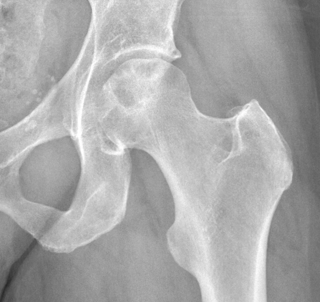
Diagnosis combines medical history, clinical examination, and imaging.
Your doctor may ask about your symptoms, previous injuries, steroid exposure, alcohol use, and relevant medical conditions.
The examination may include walking assessment, hip range of motion, and evaluation of pain during movement.
Why MRI is important
X-rays can show established bone changes and help identify collapse, but they may not detect early AVN.
MRI is particularly useful when osteonecrosis is suspected despite normal or inconclusive X-rays. It can help identify the affected area and assess the extent of disease.
CT may be used in selected cases to clarify structural changes, including suspected fracture or collapse.
The choice of imaging depends on the clinical findings and the question that needs to be answered. (Cheng et al., 2026)
Can Physiotherapy Help Avascular Necrosis of the Hip?
Physiotherapy can help maintain appropriate movement, strength, balance, and daily function. However, it does not restore dead bone tissue or reverse femoral head collapse.
Treatment must be coordinated with the orthopedic team and adapted to the stage of the disease.
1. Protect the affected hip
If the bone is structurally vulnerable or weight-bearing causes pain, the treating clinician may recommend reducing the load through the affected leg or using a walking aid.
Do not independently begin impact exercise, heavy lower-limb strengthening, or a walking program designed to push through pain if AVN is suspected or confirmed.
Appropriate loading depends on the imaging findings and the stability of the femoral head.
2. Maintain comfortable movement
When permitted by the treating team, gentle, pain-limited movement may help maintain available range and reduce unnecessary stiffness.
Avoid aggressive stretching or repeatedly forcing the hip into painful positions.
The aim is to maintain useful movement without adding unnecessary stress to the affected joint.
3. Maintain strength and function
A physiotherapist may prescribe suitable exercises for the trunk and lower limbs while respecting any weight-bearing restrictions.
The choice of exercise depends on the affected hip, symptoms, stage of disease, and whether surgery is planned.
4. Support rehabilitation after surgery
Some people with early-stage AVN may be considered for joint-preserving procedures such as core decompression.
More advanced disease with collapse may require hip replacement, depending on the individual assessment.
After surgery, physiotherapy can support safe transfers, walking with an appropriate aid, gait retraining, strength, and gradual return to daily activities.
The surgeon’s instructions determine when and how weight-bearing can progress. (Zhao et al., 2020)
Can Early Treatment Prevent Hip Joint Collapse?
Early diagnosis creates an opportunity to discuss treatment options before substantial structural damage occurs. However, early diagnosis does not guarantee that the femoral head can be saved.
For selected patients with pre-collapse disease, an orthopedic specialist may consider joint-preserving treatment.
Core decompression is one option, although its suitability and likely benefit depend on factors such as disease stage and lesion characteristics.
A meta-analysis comparing core decompression with conservative treatment found that outcomes varied according to the treatment and disease stage.
The evidence does not support assuming that one procedure is appropriate for every patient. (Hong et al., 2015)
Physiotherapy remains supportive rather than a substitute for orthopedic decision-making. If AVN is suspected, timely assessment is more important than attempting to manage the problem through exercise alone.
Common Mistakes to Avoid if You Suspect Hip AVN
Assuming mild pain means mild disease
The intensity of pain does not reliably indicate the amount of bone damage.
Early AVN can be painless, while other conditions may cause severe pain without osteonecrosis.
Continuing high-impact exercise through pain
Running, jumping, and heavy loading may be unsuitable when the femoral head is vulnerable.
Ask your treating clinician which activities and loading levels are appropriate.
Relying on an X-ray alone
A normal X-ray does not exclude early AVN. If clinical suspicion remains, a doctor may consider MRI.
Treating every symptom with stretching
Stretching cannot restore the blood supply to dead bone. Exercise should be selected to support safe function and should not delay medical investigation.
Myth vs Fact: Hip Avascular Necrosis
MYTH: Early AVN always causes severe hip pain.
FACT: Early AVN may cause mild discomfort or no symptoms. Pain intensity alone cannot determine the amount of bone damage.
MYTH: A normal X-ray rules out AVN.
FACT: Early disease may not be visible on X-rays. MRI can help detect osteonecrosis when clinical suspicion remains.
MYTH: Physiotherapy can reverse dead bone tissue.
FACT: Physiotherapy supports appropriate movement, strength, and function. It cannot restore dead bone tissue or guarantee prevention of femoral head collapse.
MYTH: Everyone with AVN should follow the same exercise plan.
FACT: Exercise and weight-bearing recommendations depend on the disease stage, bone stability, symptoms, and medical or surgical treatment plan.
When Should You Seek Medical Attention?
Arrange a timely medical assessment if you have persistent unexplained groin or hip pain, a new limp, worsening weight-bearing discomfort, or relevant risk factors for osteonecrosis.
Seek urgent medical attention if you suddenly cannot bear weight, develop severe hip pain after an injury, or experience rapidly worsening symptoms.
A hot or swollen joint accompanied by fever or feeling generally unwell also requires urgent assessment.
If AVN has already been diagnosed, attend recommended follow-up appointments even if your pain is mild.
The next steps depend on the imaging findings, the size and location of the lesion, and whether structural collapse has occurred.
Final Thoughts: Recognize the Signs Early
Avascular necrosis of the hip may begin with subtle groin pain, discomfort during walking, stiffness, or no symptoms at all. Waiting for severe pain is not a reliable way to judge the condition because the extent of bone damage cannot be determined by symptoms alone.
If you have persistent unexplained hip pain or relevant risk factors, seek medical assessment. Physiotherapy can support safe movement and rehabilitation, but protecting the joint and selecting appropriate treatment require stage-specific medical guidance.
Conclusion
Avascular necrosis of the hip can begin with subtle groin pain, discomfort during walking, stiffness, or no symptoms at all.
As the affected bone weakens, the femoral head may collapse and secondary arthritis can develop.
Recognizing risk factors and obtaining timely medical assessment are important because treatment options depend heavily on the stage of the disease.
From a physiotherapy perspective, the priority is to protect the affected hip, maintain appropriate function, and support rehabilitation under specialist guidance.
Persistent unexplained hip pain should not be dismissed simply because it is mild. Early assessment can help clarify the cause and allow appropriate treatment decisions before further damage occurs.
Frequently Asked Questions
1. Can avascular necrosis of the hip heal on its own?
Not reliably. Some small lesions may remain stable, while others progress. Management depends on the extent of affected bone and the disease stage. Medical assessment is important even when symptoms are mild.
2. Can walking make hip avascular necrosis worse?
The effect of walking depends on the condition of the affected bone. Some people need protected weight-bearing or activity modification. Follow your clinician’s advice rather than applying a universal walking or exercise recommendation.
3. Is hip avascular necrosis the same as arthritis?
No. AVN involves damage and death of bone tissue associated with impaired blood supply. If the femoral head collapses, secondary osteoarthritis may develop, but the two conditions are not the same.
4. Can physiotherapy prevent hip joint collapse?
Physiotherapy alone cannot guarantee prevention of collapse or restore dead bone tissue. It supports safe movement and rehabilitation alongside appropriate medical or surgical management.
5. Which scan detects early avascular necrosis?
MRI is particularly useful for detecting early AVN when X-rays are normal or inconclusive. A clinician determines which imaging tests are appropriate.
Stay tuned with us for more health related topics.
Follow us on LinkedIn and Instagram for more.
More Read


Medical Disclaimer
This article is intended for general educational and informational purposes only. It does not replace a medical consultation, individual assessment, or diagnosis. Hip pain can have several causes, and exercise recommendations that are appropriate for one person may not be suitable for another.
If you suspect avascular necrosis, consult a qualified healthcare professional for assessment and appropriate imaging when indicated. Seek urgent medical attention if you suddenly cannot bear weight, develop severe hip pain after an injury, or experience a hot or swollen joint with fever.


