


If your knee repeatedly clicks, snaps, locks, swells, or hurts on the outer side, you may be dealing with something more unusual than an ordinary meniscus tear.
One possibility is a discoid meniscus, a structural variation in which the meniscus is wider and more disc-shaped than the normal crescent-shaped structure.
Quick Answer: What Is a Discoid Meniscus?
A discoid meniscus is an unusually wide, disc-shaped meniscus, most often found on the outer side of the knee. Unlike a typical crescent-shaped meniscus, it covers a larger portion of the knee joint.
Not everyone with a discoid meniscus has a problem. Many people have no symptoms and never need treatment. When symptoms occur, they may include outer knee pain, clicking, snapping, swelling, catching or locking.
The interesting part is that having a discoid meniscus does not automatically mean you have a knee problem.
Many people have one for years without symptoms and discover it incidentally on an MRI.
The trouble usually begins when the unusually shaped meniscus becomes unstable, develops a tear, or starts interfering with normal knee movement.
From a physiotherapy perspective, this distinction matters enormously.
I would never want a patient to see the words “discoid meniscus” on an MRI report and immediately assume that surgery is necessary.
The goal is to understand whether the anatomy is actually causing your symptoms, whether the meniscus is torn or unstable, and how well your knee is functioning.
Recent reviews and expert consensus increasingly emphasize preserving as much functional meniscal tissue as possible when treatment is required. (Indian Journal of Orthopaedics)
Key Takeaways
What you should know about discoid meniscus
A discoid meniscus is an unusually broad, disc-shaped meniscus, most commonly affecting the outer side of the knee.
Having a discoid meniscus does not automatically mean you have a knee injury. Many people have no symptoms.
Pain, recurrent swelling, clicking, catching, snapping or locking may indicate that the meniscus has become symptomatic or unstable.
MRI can help identify a discoid meniscus and associated tears, but imaging findings should always be interpreted alongside symptoms and clinical examination.
Physiotherapy can improve knee strength, mobility, balance and movement control, although it cannot change the anatomical shape of the meniscus.
When surgery is necessary, modern treatment generally aims to preserve as much healthy meniscal tissue as possible rather than removing the entire meniscus.
An asymptomatic discoid meniscus usually does not require preventive surgery. Treat the symptoms and function, not the MRI report alone.
What Is a Discoid Meniscus?
A meniscus is a tough, crescent-shaped piece of fibrocartilage between the thigh bone and shin bone.
Each knee has a medial meniscus on the inner side and a lateral meniscus on the outer side.
A normal meniscus helps distribute load, improve joint stability, assist lubrication and protect the articular cartilage.
A discoid meniscus is different because it is broader and more disc-like instead of having the usual C-shaped appearance.
The lateral meniscus is affected far more often than the medial meniscus.
This is why you will often see the condition referred to as a discoid lateral meniscus (DLM).
Reported prevalence varies substantially according to population and diagnostic method.
Expert consensus estimates approximately 3%-5% in Western populations and around 10.9%-16.6% in Asian populations. (BMC MSK Disorder)
One particularly interesting finding is that discoid menisci may occur in both knees.
A systematic review found bilateral involvement was especially common in East Asian populations, although reported rates varied considerably between studies.
That does not mean both knees need treatment.
In fact, an asymptomatic discoid meniscus generally does not require preventive surgery.
Why Does the Meniscus Become “Discoid”?
This is one area where the medical literature is more complicated than many online articles suggest.
A discoid meniscus is generally regarded as a congenital or developmental anatomical variant, but its precise developmental mechanism remains debated.
Genetics may also contribute.
Importantly, it is not something you develop simply because you played football, ran too much, squatted incorrectly, or exercised at the gym.
What physical activity can do is expose a vulnerable meniscus to repeated loading.
The abnormal shape may alter how forces are distributed through the knee.
In addition, some discoid menisci have abnormal peripheral attachments or instability.
These factors can make the structure more susceptible to tearing.
This is an important distinction:
Exercise does not necessarily create the discoid meniscus. It may reveal a problem that was already present.
The Three Main Types of Discoid Meniscus
Orthopaedic literature commonly describes discoid lateral menisci according to the Watanabe classification.
Complete discoid meniscus
This type covers most or essentially the entire lateral tibial plateau.
Because there is more meniscal tissue than usual, it may have a pronounced disc-like appearance.
Incomplete discoid meniscus
The meniscus is still wider than normal but does not cover the entire lateral tibial plateau.
It retains more of the normal crescent-like configuration than the complete type.
Wrisberg-type discoid meniscus
This is particularly interesting.
The meniscus may have a discoid shape but lacks some of its normal posterior attachments, making it potentially unstable.
Patients may experience a characteristic snapping or popping sensation, particularly during knee movement.
The type alone, however, does not tell us whether someone needs treatment.
Symptoms, tears, instability, cartilage status, activity level and examination findings all matter.
What Does a Discoid Meniscus Feel Like?
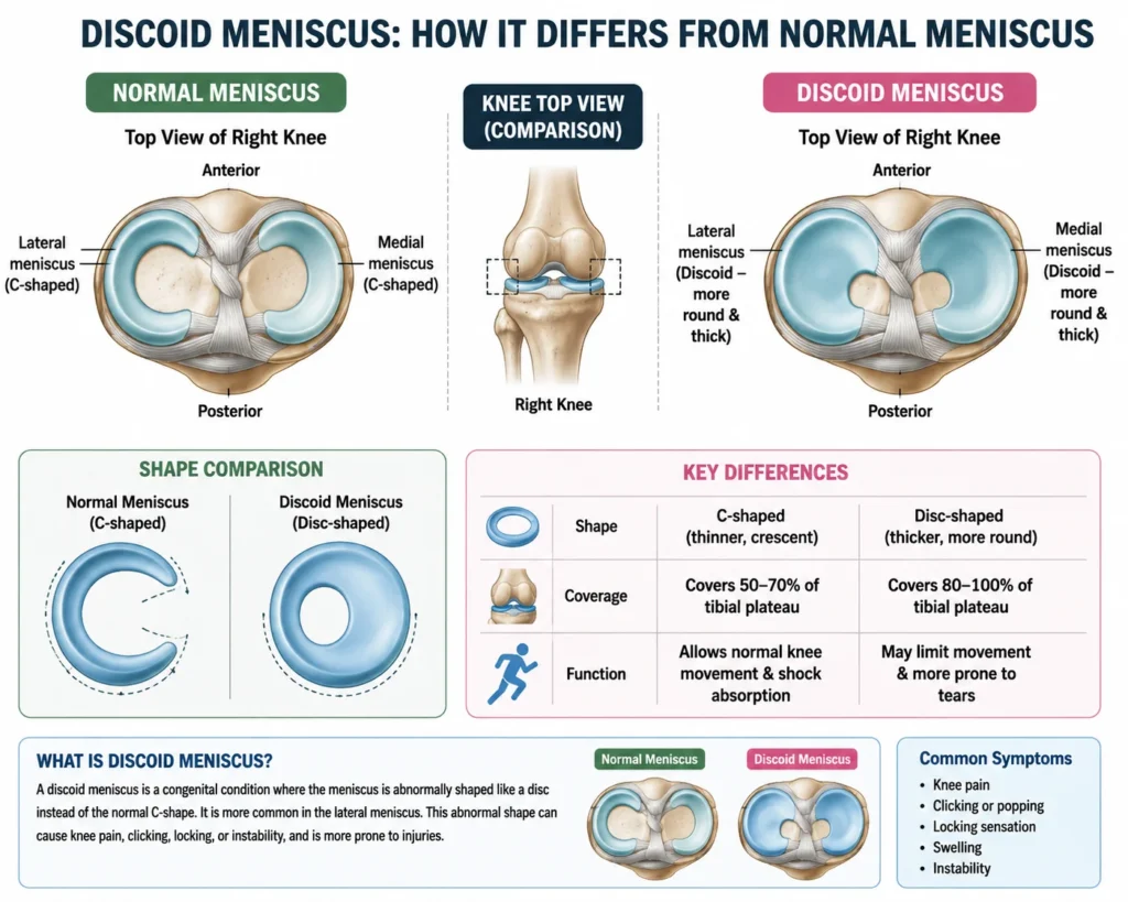
Some people feel absolutely nothing.
Others develop symptoms gradually or after a twisting injury.
Common symptoms include:
- Pain along the outer side of the knee
- Clicking or popping
- Recurrent swelling
- A sensation that the knee catches
- Locking or difficulty fully straightening the knee
- Giving way or instability
- Reduced confidence during running or sports
- Pain during squatting or twisting
- A snapping sensation during movement
The pattern is more informative than any single symptom.
For example, a painless click does not automatically mean that the meniscus is damaged.
Likewise, knee pain does not automatically mean that the discoid meniscus is responsible.
This is why an MRI finding must be interpreted alongside the history and physical examination.
One Lesser-Known Clue: The “Snapping Knee”
A snapping knee deserves more attention than it often receives.
In some children and adolescents, a discoid meniscus may produce a reproducible snapping sensation when the knee moves between flexion and extension.
This can happen because the abnormal meniscus moves abnormally within the joint.
However, a snap without pain, swelling, locking or functional limitation does not automatically require surgery.
Historical literature and modern consensus both support observation in appropriately selected asymptomatic or minimally symptomatic patients.
As a physiotherapist, I would therefore ask:
Is the clicking merely a sound, or does it represent a mechanical problem?
That distinction can prevent unnecessary treatment.
Discoid Meniscus vs Meniscus Tear: They Are Not the Same Thing
This is one of the most important points to understand.
A discoid meniscus is primarily an anatomical shape variation.
A meniscus tear is an injury or structural disruption of meniscal tissue.
You can have:
- A discoid meniscus without a tear
- A discoid meniscus with a tear
- A discoid meniscus with instability
- A discoid meniscus with both tearing and instability
The presence of a discoid meniscus therefore does not automatically mean that the meniscus is torn.
Recent systematic research also suggests that tear patterns may differ according to age.
Adults appear more likely to present with complex tears, while horizontal tear patterns are frequently reported in children. (Wiley)
This is one reason I would avoid making treatment decisions based solely on the phrase “discoid meniscus” in an MRI report.
How Is Discoid Meniscus Diagnosed?
Your clinician will usually combine three things:
Clinical examination
A physiotherapist or orthopaedic clinician may assess:
- Knee range of motion
- Joint-line tenderness
- Swelling
- Walking pattern
- Squatting mechanics
- Single-leg control
- Ligament stability
- Meniscal provocation tests
- Quadriceps and hip strength
- Mechanical symptoms
The physical examination is useful, but it cannot always distinguish a discoid meniscus from other causes of lateral knee pain.
X-ray
An X-ray cannot directly show the meniscus as well as MRI can.
However, certain indirect findings may raise suspicion and X-rays can also help identify other causes of knee pain.
MRI
MRI is generally the most useful non-invasive imaging method for confirming a discoid meniscus and identifying associated tears, cartilage abnormalities and other internal knee problems.
One fascinating radiological clue is the “bow-tie” appearance on sagittal MRI images.
A normal meniscus appears on sequential sagittal slices as separate anterior and posterior components.
A discoid meniscus can remain continuous across several slices, producing the characteristic appearance.
Older MRI research established useful measurement criteria for identifying discoid lateral menisci, including meniscal width and the proportion of the tibial plateau covered by the meniscus. (OJSM)
Can You Have a Discoid Meniscus Without Knowing It?
Absolutely.
This is probably the most reassuring fact about the condition.
A discoid meniscus can remain completely asymptomatic.
The knee can adapt to the anatomical variation, and many people function normally without ever needing treatment.
Current expert guidance specifically recommends conservative management for asymptomatic discoid lateral menisci rather than prophylactic surgery. (Reviews in MSK Medicine)
This is also why I would not recommend trying to “fix” an MRI finding simply because it exists.
Treat the patient, not the scan.
When Does a Discoid Meniscus Need Treatment?
Treatment depends primarily on symptoms and mechanical status.
Conservative management may be appropriate when:
- There is no significant pain
- There is no recurrent swelling
- The knee is stable
- There is no significant locking
- Daily activities remain comfortable
- Sports participation is not meaningfully restricted
Surgery becomes more relevant when symptoms persist despite appropriate conservative care, or when there is a significant tear or instability producing mechanical symptoms.
The Chinese Society of Sports Medicine consensus recommends considering arthroscopic treatment for symptomatic discoid lateral meniscus with significant functional impact, particularly when tearing or peripheral instability is present. (PMC)
Physiotherapy Treatment for Discoid Meniscus
Physiotherapy is not designed to change a discoid meniscus back into a normal-shaped meniscus.
Instead, the aim is to make the knee stronger, better controlled and more tolerant of load, while reducing irritation.
Step 1: Reduce the aggravating load
If your knee repeatedly becomes painful after deep squatting, pivoting or high-volume running, temporarily reducing those loads may be more useful than complete rest.
This is where physiotherapy differs from simply saying, “Stop exercising.”
I prefer load modification rather than fear-based avoidance.
For example, a runner may temporarily reduce mileage while maintaining cycling, walking or appropriately selected strengthening exercises if these are comfortable.
Step 2: Restore full knee movement
A painful or swollen knee may lose extension.
That matters.
Reduced extension can alter walking mechanics and place additional demands on surrounding muscles.
A physiotherapist may use gentle mobility work, active range-of-motion exercises and progressive loading to restore normal movement.
Step 3: Rebuild quadriceps strength
The quadriceps are crucial for controlling the knee during walking, stairs, landing and sport.
Depending on symptoms, rehabilitation may progress through exercises such as:
- Straight-leg raises
- Sit-to-stand movements
- Mini squats
- Step-ups
- Leg presses
- Split squats
- Controlled single-leg exercises
The exact exercise selection should be individualized.
Pain during an exercise is not automatically dangerous, but persistent swelling or worsening mechanical symptoms is a reason to reassess the program.
Step 4: Do not ignore the hip
One lesser-discussed aspect of knee rehabilitation is proximal control.
Weakness or poor coordination around the hip can influence lower-limb movement during running, landing and single-leg tasks.
Therefore, a complete program may include:
- Hip abductor strengthening
- Gluteal strengthening
- Single-leg balance
- Pelvic control exercises
- Movement retraining
This does not “cure” the discoid anatomy. It improves the overall system around the knee.
What Happens If Surgery Is Recommended?
The modern philosophy is increasingly meniscus preservation.
Instead of simply removing as much meniscal tissue as possible, surgeons may reshape the discoid meniscus into a more normal configuration, commonly called saucerization or meniscoplasty, while preserving a stable peripheral rim.
If the meniscus is unstable or has a repairable tear, repair may be performed at the same time.
The objective is not simply to make the MRI look normal.
The objective is to preserve as much useful meniscal tissue as possible.
That is important because the meniscus contributes substantially to load distribution and protection of articular cartilage.
Why Removing Too Much Meniscus Can Be a Problem
Historically, total meniscectomy was used more frequently.
We now understand the long-term consequences much better.
Long-term research suggests that subtotal or total meniscectomy is associated with greater degenerative change, which is one reason contemporary treatment favors preservation whenever technically possible.
A particularly striking 2025 study following athletes treated for discoid meniscus reported considerably better long-term osteoarthritis outcomes in the meniscus-preserving group compared with subtotal or total meniscectomy. (Wolters Kluwer)
This does not mean every discoid meniscus should be preserved at all costs.
Some tissue may be irreparable or nonfunctional.
But the principle is clear:
If healthy, stable meniscal tissue can safely be preserved, preserving it matters.
Can a Discoid Meniscus Cause Knee Arthritis?
Potentially, yes.
But it is inaccurate to tell someone that a discoid meniscus automatically causes arthritis.
Risk appears to depend on several factors, including tears, instability, meniscal tissue remaining after surgery, alignment, cartilage health and duration of symptoms.
One study found that prolonged symptoms, greater discoid meniscus thickness and horizontal tears were associated with increased risk of radiographic osteoarthritis after partial meniscectomy. (Science Direct )
A horizontal tear was the strongest predictor in that analysis.
Another long-term study of young patients after partial meniscectomy reported progression of lateral-compartment radiographic arthritis in a substantial proportion over approximately 10 years. (NCBI)
These findings are exactly why long-term thinking matters.
The goal is not merely:
“How quickly can we stop the knee from hurting?”
It is also:
“How can we preserve the knee for the next 20 or 30 years?”
Rehabilitation After Discoid Meniscus Surgery
Rehabilitation varies depending on whether you had saucerization alone, meniscal repair, or another procedure.
That distinction is crucial.
Someone who undergoes a simple reshaping procedure may progress differently from someone whose meniscus has been repaired and needs biological healing time.
Early rehabilitation commonly focuses on:
- Controlling swelling
- Restoring knee extension
- Gradually restoring flexion
- Re-establishing quadriceps activation
- Normalizing walking
- Progressive strength training
Later stages emphasize:
- Single-leg strength
- Balance
- Dynamic control
- Running preparation
- Landing mechanics
- Sport-specific drills
Research has shown that physical deficits can persist during recovery.
A 2024 study found knee extensor weakness and altered gait mechanics during the earlier months following saucerization and repair, with some measures improving by 12 months. (Frontiers in Medicine)
This is an important reminder not to judge recovery solely by pain.
Pain may disappear before strength and movement quality are fully restored.
How Long Does Recovery Take?
There is no single recovery timeline.
After a procedure involving only reshaping, rehabilitation may progress relatively quickly.
After meniscal repair, the surgeon may impose more restrictions to protect healing tissue.
Return to running and sport should therefore be based on criteria rather than a calendar date alone.
Before returning to higher-level activity, I would want to see:
- Minimal or no swelling
- Full or near-full range of motion
- Good quadriceps strength
- Good single-leg control
- Ability to perform functional movements confidently
- No significant catching or locking
- Progressive tolerance to impact
Returning to sport simply because “three months have passed” is not a sufficient rehabilitation strategy.
What Should You Avoid With a Symptomatic Discoid Meniscus?
You do not necessarily have to stop all physical activity.
Instead, be cautious with activities that consistently provoke symptoms.
During an irritated phase, temporarily reduce:
- Deep loaded squats if painful
- Repeated pivoting
- Sudden direction changes
- High-volume jumping
- Aggressive twisting
- Long runs that repeatedly trigger swelling
- Training through mechanical locking
One of the biggest mistakes I see is alternating between complete rest and aggressive exercise.
A better approach is usually progressive loading.
If walking is comfortable but running causes swelling, maintain comfortable activity while temporarily reducing running load.
Lesser-Known Things That Can Help
Watch the swelling, not just the pain
A knee that becomes repeatedly swollen after activity deserves attention even if the pain is mild.
Swelling can be a more useful indicator of joint irritability than pain alone.
Track mechanical symptoms
Write down when clicking, catching or locking occurs.
Does it happen while extending the knee?
After sitting?
During squatting?
When turning?
This information can help your clinician understand whether the symptoms are mechanically reproducible.
Do not self-test the knee repeatedly
If you have discovered that twisting your knee produces a click, repeatedly performing the movement to “check whether it is still there” is not useful.
You may simply irritate the joint.
Strength matters even when pain improves
Feeling better does not necessarily mean the knee has regained its pre-injury capacity.
Progressive strengthening remains important during recovery.
Do not assume the other knee needs surgery
Bilateral discoid menisci are common, but an asymptomatic opposite knee generally should not undergo prophylactic surgery simply because imaging reveals the same anatomy.
When Should You See an Orthopaedic Specialist?
Seek professional assessment if you develop:
- Recurrent knee locking
- Inability to fully straighten the knee
- Repeated swelling
- Persistent lateral knee pain
- Knee instability
- Symptoms following a twisting injury
- Difficulty returning to normal activity
- Mechanical symptoms that are progressively worsening
An acutely locked knee deserves particular attention because a displaced meniscal fragment or other intra-articular problem may be involved.
Can You Prevent Discoid Meniscus?
You cannot prevent the anatomical shape itself.
You were born with the variation or developed it during growth.
But you can potentially reduce the likelihood of aggravating it by managing training loads, maintaining lower-limb strength, responding early to persistent swelling and avoiding repeatedly pushing through mechanical symptoms.
For athletes, sensible progression is particularly important.
The aim is not to become afraid of movement.
It is to develop a knee capable of handling the amount of movement you are asking it to perform.
The Bottom Line
A discoid meniscus is not automatically a damaged meniscus.
It is an unusual meniscal shape, most commonly affecting the lateral meniscus.
Many people have this anatomical variation without symptoms and never require treatment.
Problems arise when the discoid meniscus becomes torn, unstable or mechanically symptomatic.
The modern approach is therefore much more nuanced than the old idea of simply removing the meniscus.
When surgery is necessary, contemporary treatment increasingly emphasizes saucerization, repair and preservation of functional meniscal tissue whenever appropriate.
From a physiotherapy perspective, I would focus on the person rather than the MRI alone.
A strong, well-controlled knee with no swelling, locking or functional limitation may need observation rather than intervention.
A knee that repeatedly swells, locks or becomes unstable deserves a proper assessment.
And if surgery is required, rehabilitation should not end when the pain disappears.
Restoring strength, movement quality, confidence and load tolerance is what helps you return to everyday life or sport safely.
The most important message I would leave you with is simple:
A discoid meniscus is an anatomical variation.
A symptomatic discoid meniscus is a clinical problem.
They are not the same thing.
Frequently Asked Questions
Clear answers to common questions about symptoms, treatment, surgery and recovery.
A discoid meniscus is an unusually wide, disc-shaped meniscus rather than the typical crescent-shaped meniscus. It most commonly affects the lateral meniscus on the outer side of the knee. Some people have a discoid meniscus without experiencing any symptoms.
Symptoms may include pain on the outer side of the knee, clicking, snapping, recurrent swelling, catching, stiffness, instability or difficulty fully straightening the knee. Some people remain completely symptom-free despite having a discoid meniscus.
No. An asymptomatic discoid meniscus generally does not require surgery. Treatment depends on symptoms, associated tears, instability, mechanical locking and how much the condition interferes with daily activities or sport.
Yes. Depending on the condition, non-surgical management may include activity modification, progressive strengthening, mobility exercises, balance training and physiotherapy. However, persistent locking, significant tearing or instability may require assessment by an orthopaedic specialist.
Physiotherapy cannot change the shape of a discoid meniscus, but it can improve knee strength, range of motion, balance, movement control and tolerance to physical activity. Physiotherapy is also an important part of rehabilitation after appropriate discoid meniscus surgery.
Depending on symptoms and the stage of rehabilitation, exercises may include quadriceps strengthening, straight-leg raises, controlled squats, step-ups, hip strengthening, balance exercises and progressive single-leg movements. Exercises should be selected according to the individual’s symptoms and clinical assessment.
Diagnosis usually involves a clinical examination and imaging. X-rays may help rule out other knee problems, while MRI is particularly useful for identifying the discoid shape and detecting associated meniscal tears, instability or cartilage abnormalities.
Recovery varies depending on the procedure. Recovery after reshaping alone may progress differently from recovery after meniscal repair. Rehabilitation commonly focuses first on swelling control, range of motion and muscle activation before progressing to strength, balance, running and sport-specific activities.
A discoid meniscus does not automatically cause arthritis. However, associated tears, instability, cartilage damage and loss of meniscal tissue may increase long-term degenerative risk. This is one reason modern treatment often emphasizes preserving functional meniscal tissue whenever possible.
Seek professional assessment if you have persistent outer knee pain, recurrent swelling, repeated catching or locking, instability, difficulty fully straightening the knee, or symptoms that interfere with walking, exercise or sports. A suddenly locked knee should be assessed promptly.
Stay tuned with us for more health related topics.
Follow us on LinkedIn and Instagram for more.
More Read


Medical Disclaimer!
This article has been reviewed and written under the guidance of our Head Physiotherapist, Dr. Kruti Raj (PT, MUHS,CPT,CMPT). The information shared is intended for educational purposes only and should not be considered a substitute for personalized medical advice, diagnosis, or treatment.
Please consult us or any other qualified healthcare professional before beginning any exercise program, especially if you are experiencing pain, recovering from injury, or managing a medical condition.


