


Femoral head osteonecrosis occurs when the bone at the top of the thigh bone loses its normal blood supply, causing bone tissue to die.
Quick Answer: Femoral Head Osteonecrosis
Femoral head osteonecrosis, also called avascular necrosis (AVN) of the hip, occurs when reduced blood supply damages the bone at the top of the thigh bone. Common symptoms include deep groin pain, hip stiffness, discomfort during walking, and difficulty bearing weight.
The condition progresses through stages, from early bone changes to possible femoral head collapse and secondary arthritis. MRI can help detect early disease when X-rays appear normal.
Also known as avascular necrosis (AVN) of the hip, it can cause deep groin pain, stiffness, and difficulty walking. In its early stages, the condition may cause few symptoms and remain invisible on routine X-rays.
Without appropriate management, the weakened bone may develop a fracture beneath the joint surface and eventually collapse.
Early diagnosis matters because selected patients may be eligible for treatments designed to preserve the natural hip before substantial collapse occurs.
Key Takeaways
- Femoral head osteonecrosis develops when reduced blood supply damages bone tissue in the hip joint.
- Risk factors include corticosteroid exposure, heavy alcohol consumption, certain blood disorders, and hip trauma.
- Early disease may cause few symptoms and may not be visible on routine X-rays.
- MRI can help detect osteonecrosis before the femoral head collapses.
- Treatment depends on disease stage, lesion size, symptoms, and individual health factors.
- Core decompression and other joint-preserving procedures may be considered in selected patients before substantial collapse.
- Total hip replacement may be appropriate when advanced damage causes significant pain and disability.
- Physiotherapy can support safe movement, strength, walking, and postoperative recovery, but it cannot reverse dead bone by itself.
What Is Femoral Head Osteonecrosis?
The femoral head is the rounded upper end of the thigh bone that fits into the socket of the pelvis. Its smooth cartilage-covered surface allows the hip to move during walking, sitting, and climbing stairs.
Osteonecrosis develops when blood flow to part of the bone is disrupted.
As bone cells die, the affected area can weaken and become less able to withstand everyday forces.
A fracture beneath the joint surface may then cause the femoral head to flatten or collapse.
This structural damage can alter how forces travel through the hip and contribute to secondary osteoarthritis.
Osteonecrosis may affect one or both hips. Some people have no symptoms initially, while others develop progressive pain and mobility limitations. (Gorge et al., 2022)
What Causes Femoral Head Osteonecrosis?
Several factors can interfere with the blood supply or health of the femoral head.
1. Corticosteroid use
Prolonged or high-dose corticosteroid treatment is associated with an increased risk of osteonecrosis.
However, not everyone taking these medicines develops the condition.
Never stop prescribed steroids without medical guidance.
2. Heavy alcohol consumption
Heavy alcohol use is associated with osteonecrosis and may affect bone-cell function and blood supply.
Risk varies between individuals and can be influenced by other medical factors.
3. Hip injuries
A hip dislocation or certain fractures can damage the blood vessels supplying the femoral head.
Osteonecrosis may appear after the initial injury, sometimes months or years later.
4. Blood and autoimmune disorders
Sickle cell disease and certain clotting or blood disorders can interfere with circulation.
Some autoimmune conditions are also associated with osteonecrosis, sometimes alongside the effects of their treatment.
5. Other medical conditions
Osteonecrosis has been reported in association with organ transplantation, some cancer treatments, and other conditions affecting circulation or bone health.
In some cases, no clear cause is identified.
Having a risk factor does not confirm the diagnosis.
Persistent hip or groin pain should be investigated rather than automatically attributed to a previous injury, medicine, or lifestyle factor. (Konarski et al., 2023)
Six Symptoms of Femoral Head Osteonecrosis
Symptoms depend on the size and location of the affected area and whether structural damage has developed.
1. Deep groin pain
Pain may develop deep in the groin and sometimes spread towards the thigh or buttock. It can be mistaken for a muscle strain or another hip problem.
2. Pain during weight-bearing
Walking, standing for long periods, and climbing stairs may become uncomfortable. As the condition progresses, everyday activities may become more difficult.
3. Pain during hip movement
Turning the leg, getting into a car, or bending the hip may provoke discomfort. Reduced hip rotation can develop as the joint becomes more affected.
4. Limping
Pain may cause a person to shorten their stride or shift weight towards the other leg. A new or worsening limp deserves assessment.
5. Stiffness and reduced mobility
The hip may feel less flexible, particularly during rotation. Pain-related guarding and later joint changes can contribute to restricted movement.
6. Pain at rest or at night
More advanced disease may cause discomfort even while resting or sleeping. However, symptom intensity alone cannot determine the stage.
Important: Early osteonecrosis can be painless. People with relevant risk factors and unexplained hip pain should discuss appropriate assessment with a healthcare professional.
What Are the Four Stages of Femoral Head Osteonecrosis?
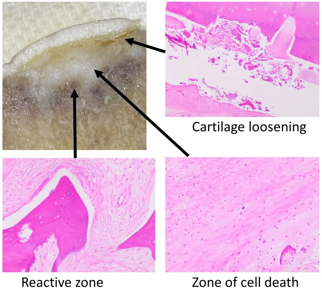
The Association Research Circulation Osseous (ARCO) system describes four stages based on imaging and structural changes.
Stage I: Early disease
X-rays may appear normal, but MRI identifies changes consistent with osteonecrosis. The femoral head has not collapsed.
Treatment consideration: The specialist assesses lesion size, location, symptoms, and the risk of progression.
Stage II: Bone changes without fracture or collapse
Imaging may show sclerosis, cyst-like changes, or other bone abnormalities, but there is no subchondral fracture or femoral head collapse.
Treatment consideration: Selected patients may be considered for joint-preserving treatment.
Stage III: Subchondral fracture
A fracture develops in the bone just beneath the joint surface. Flattening or collapse may also occur.
The ARCO system distinguishes early collapse, with depression of 2 mm or less, from more advanced collapse greater than 2 mm.
Treatment consideration: Joint preservation may still be possible in selected cases, but the outlook depends on the extent of damage.
Stage IV: Secondary osteoarthritis
The femoral head has undergone structural damage, and degenerative changes affect the hip joint. Joint-space narrowing and changes involving the socket may develop.
Treatment consideration: Total hip replacement may be considered when pain and functional limitations are substantial.
These stages do not predict an identical course for every patient. Accurate staging, lesion characteristics, and clinical assessment help guide treatment. (Cheng et al., 2026)
How Is Osteonecrosis Diagnosed?
Clinical assessment
A clinician reviews the pain pattern, walking ability, hip movement, previous injuries, medication history, and relevant medical conditions.
Physical examination helps assess function but cannot confirm osteonecrosis on its own.
X-rays
X-rays can identify later bone changes, collapse, and arthritis. However, early disease may not be visible.
MRI
MRI is particularly useful when osteonecrosis is suspected despite normal or inconclusive X-rays.
It can identify early bone changes and help determine the size and location of the affected area.
CT scans
CT may be useful when a specialist needs a clearer assessment of a suspected fracture beneath the joint surface or the extent of bone collapse.
Other conditions, including hip osteoarthritis, bone marrow oedema syndrome, and stress or insufficiency fractures, can cause similar symptoms.
Diagnosis should therefore combine clinical information with appropriate imaging. (Expert Panel on Musculoskeletal Imaging, 2022)
What Are the Treatment Options?
Treatment depends on the stage, lesion size and location, symptoms, age, activity needs, and overall health.
1. Activity modification
Reducing high-impact activity may help manage symptoms. A clinician may recommend crutches or reduced weight-bearing in selected cases.
These measures can improve comfort, but they do not reliably restore dead bone or guarantee that collapse will be prevented.
2. Core decompression
Core decompression is a surgical procedure considered for selected patients, particularly before the femoral head collapses.
The surgeon creates channels in the affected bone to encourage a healing response.
Bone grafting or other additional procedures may be considered in specific cases. Results vary, and core decompression is not suitable for every patient.
3. Bone grafting and other joint-preserving procedures
Selected patients may be offered bone grafting, vascularized bone graft procedures, or osteotomy.
These aim to support the affected region or redistribute forces through the joint.
Suitability depends on the lesion and the amount of structural damage. Evidence for different procedures varies, so decisions should be individualized.
4. Total hip replacement
When substantial collapse and secondary arthritis cause persistent pain and disability, total hip replacement may provide more reliable relief.
The damaged joint surfaces are replaced with prosthetic components.
Although it is a major operation, it can improve mobility and quality of life when preserving the original joint is unlikely to succeed.
Evidence reviews show that outcomes from core decompression vary with disease stage and the procedure used. It should not be presented as a guaranteed way to prevent hip replacement. (Hua et al., 2019)
How Does Physiotherapy Help?
Physiotherapy supports movement, strength, walking ability, and functional recovery.
However, exercise cannot restore dead bone or reverse femoral head collapse by itself. Rehabilitation should be coordinated with the orthopedic team.
1. Safe walking and load management
A physiotherapist can teach patients to use crutches or other walking aids when reduced weight-bearing is prescribed.
The appropriate amount of loading depends on the condition and any surgical treatment.
2. Maintaining hip mobility
Gentle, symptom-guided movement may help maintain mobility when approved by the treating team. Forceful stretching or movements that significantly increase pain should be avoided.
3. Restoring strength and balance
When appropriate, rehabilitation may include progressive strengthening of the hip and surrounding muscles, balance exercises, and gait retraining.
Exercise selection should reflect the stage of disease and the patient’s functional limitations.
4. Rehabilitation after surgery
Following core decompression or hip replacement, physiotherapy may help patients follow movement precautions, improve walking, and gradually rebuild strength.
Loading and exercise progression should follow the surgeon’s protocol.
5. Supporting everyday activities
Physiotherapists can help patients adapt stairs, transfers, work tasks, and other daily activities to their current abilities.
This can help maintain independence while medical treatment addresses the underlying condition.
Can Femoral Head Osteonecrosis Be Prevented From Progressing?
Progression cannot always be prevented, but early assessment can help identify treatment options before major structural damage develops.
Depending on the cause and medical circumstances, useful steps may include:
- Discussing corticosteroid risks and treatment alternatives with the prescribing clinician.
- Seeking support to reduce heavy alcohol consumption.
- Following medical advice for underlying blood or autoimmune conditions.
- Attending recommended reviews and imaging appointments.
- Avoiding high-impact activity or unapproved loading when advised by the treating team.
Do not stop medication or begin a restrictive exercise program without professional guidance.
Femoral Head Osteonecrosis: Myth vs Fact
MYTH: A normal X-ray means there is no osteonecrosis.
FACT: Early osteonecrosis may not appear on X-rays. MRI may be recommended when clinical suspicion remains.
MYTH: Physiotherapy exercises can restore dead bone.
FACT: Physiotherapy supports mobility, strength, and function, but it cannot independently restore dead bone or reverse structural collapse.
MYTH: Everyone with osteonecrosis needs a hip replacement.
FACT: Treatment varies by stage. Selected patients with early disease may be considered for joint-preserving procedures.
MYTH: Mild pain means the condition is harmless.
FACT: Symptoms do not reliably indicate the extent of bone damage. Persistent unexplained hip or groin pain deserves medical assessment.
When Should You Seek Medical Attention?
Arrange an assessment for persistent or unexplained groin or hip pain, especially if you have a history of hip trauma, corticosteroid use, heavy alcohol consumption, or a condition associated with osteonecrosis.
Seek urgent medical care if pain suddenly becomes severe, you cannot bear weight, or symptoms develop after a significant injury.
These signs may indicate a serious hip problem requiring prompt investigation.
Do not assume that mild pain means the bone is healthy, and do not rely on exercises alone to manage suspected osteonecrosis.
Final Thoughts: Protecting Your Hip Joint
Femoral head osteonecrosis can be difficult to recognize in its early stages, but timely assessment may help identify treatment options before major structural damage occurs. Persistent groin pain, stiffness, and difficulty bearing weight should not be ignored, particularly when risk factors are present.
Treatment must be tailored to the stage of the condition. Physiotherapy can support safe movement, strength, walking, and recovery after surgery, while the orthopedic team determines how best to manage the damaged bone.
Conclusion
Femoral head osteonecrosis occurs when reduced blood supply damages bone at the top of the thigh bone.
Early disease may be difficult to detect on X-rays, making MRI important when clinical suspicion remains.
Treatment ranges from activity modification and monitoring to joint-preserving surgery and total hip replacement.
Physiotherapy plays a valuable role in maintaining safe movement, improving strength, and supporting rehabilitation, but it does not replace medical treatment of the underlying bone damage.
Early diagnosis and stage-appropriate care are essential.
If persistent hip or groin pain is affecting your daily activities, seek professional assessment rather than relying on exercises alone.
Frequently Asked Questions
1. What is femoral head osteonecrosis?
Femoral head osteonecrosis is a condition in which reduced blood supply damages bone tissue at the top of the thigh bone. It can weaken the femoral head and lead to collapse or secondary arthritis.
2. What are the early symptoms of hip osteonecrosis?
Early symptoms may include deep groin pain, discomfort during weight-bearing, or pain with hip movement. Some people have no symptoms during the early stages.
3. Can an X-ray miss avascular necrosis of the hip?
Yes. Early osteonecrosis may not be visible on an X-ray. MRI can help detect early bone changes when the diagnosis is suspected.
4. Can femoral head osteonecrosis heal without surgery?
Management depends on the stage, lesion size, symptoms, and risk of collapse. Some patients may be monitored or managed without surgery, while others may benefit from joint-preserving procedures or hip replacement.
5. Can physiotherapy help with femoral head osteonecrosis?
Physiotherapy may help maintain safe mobility, improve strength, support walking, and aid postoperative recovery. It cannot independently reverse bone death or restore a collapsed femoral head.
6. When is hip replacement needed?
Hip replacement may be considered when substantial femoral head collapse or secondary arthritis causes persistent pain and functional limitations. The decision depends on the clinical findings and individual circumstances.
Stay tuned with us for more health related topics.
Follow us on LinkedIn and Instagram for more.
More Read


Medical Disclaimer
This article is intended for general educational purposes only and is not a substitute for professional medical advice, diagnosis, or treatment. Hip and groin pain can have several causes, and symptoms alone cannot confirm or exclude femoral head osteonecrosis.
Consult a qualified healthcare professional for persistent or unexplained hip pain. Seek urgent medical attention for sudden severe pain or an inability to bear weight. Do not stop prescribed medication or begin a new rehabilitation program without appropriate professional guidance.


